Nervous System Diagram

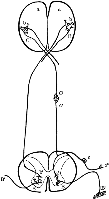
Diagram of nervous system. Labels: a, a, cortex of cerebral hemispheres; b, b, cell body and dendrites of upper motor neuron, situated in cerebral cortex; b’, axon of upper motor neuron, branching at its termination near the dendrites of lower motor neuron, situated in the ventral horn of gray matter in the spinal cord; B’, axon of lower motor neuron passing to its termination in a voluntary muscle fiber B"; C, cell body and dendrites of supper sensory neuron, situated in the medulla oblongata; C’C’, axons of upper sensory neurons, terminating in cortex; c, cell body of lower sensory neuron situated in the dorsal root ganglion; c’’’, dendrite of lower motor neuron, conducting impulses from the periphery to the central nervous system; c", long axon of lower sensory neuron, conducting impulses toward the brain; c’, short axon of lower sensory neuron, conducting impulses direct to ventral horn. (For the sake of simplicity the connection with the cerebellum are omitted.)
Galleries
Human Central Nervous SystemSource
Kimber, Diana C. Anatomy and Physiology for Nurses (New York, NY: The Macmillan Company, 1907)
Downloads
1316×2400, 197.1 KiB
561×1024, 34.1 KiB
350×640, 18.8 KiB
175×320, 8.1 KiB