Nerve Fibers

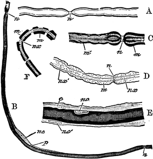
To illustrate the structure of nerve fibers. Labels: A, nerve fiber examined fresh; n, node. B, nerve fiber with axis cylinder shaded, and medulla represented by dark lines; n.c, nucleus; p, granular cell substance near the nucleus. C, more highly magnified: m, medulla; n, node. D, nerve treated with reagents to show the axis cylinder: n.x, surrounded by medulla, m. E, nerve treated with reagents to show n.c, nucleus with fine line over it representing the neurilemma, and outside this fine connective tissue, c: n.c’, nucleus lying in the fine connective tissue. F, nerve fiber deprived of its neurilemma showing medulla broken up into fragments, m, surrounding the axis cylinder, n.x.
Galleries
Human Peripheral Nervous SystemSource
Martin, H. Newell The Human Body: A Text-book of Anatomy, Physiology and Hygiene (New York: Henry Holt and Company, 1900) 244
Downloads
2313×2400, 839.7 KiB
986×1024, 131.0 KiB
616×640, 63.4 KiB
308×320, 22.3 KiB