Right Atrium and Ventricle of the Heart
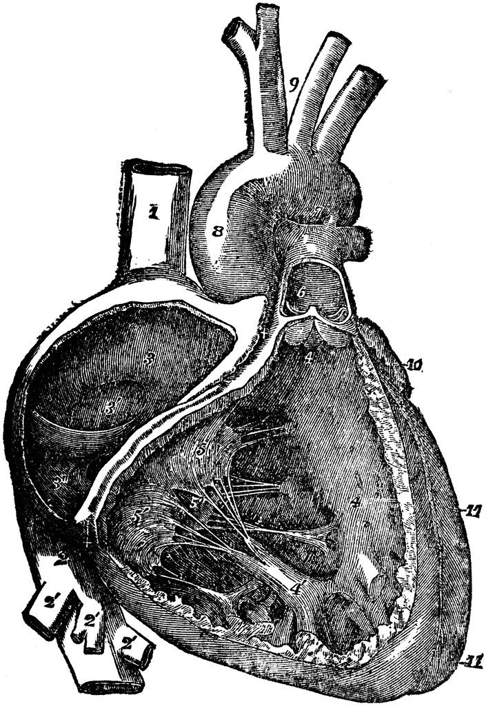
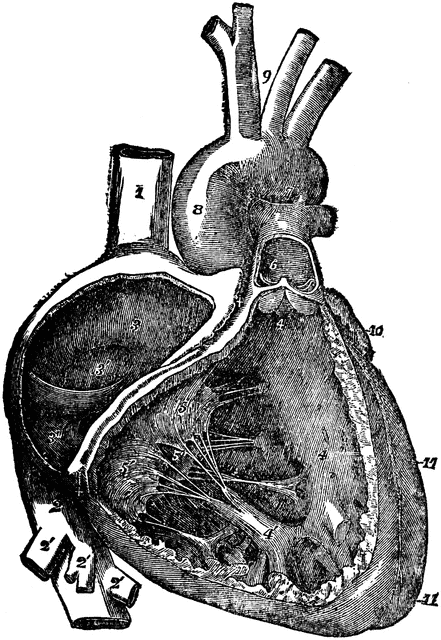
The right auricle (atrium) and ventricle of the heart opened, and a part of their right and anterior walls removed, so as to show their interior; 1/2, 1, superior vena cava; 2, inferior vena cava; 2’, hepatic veins cut short; 3, right auricle; 3’, placed in the fossa ovalis, below which is the Eustachian valve; 3", is placed close to the aperture of the coronary vein; +, +, placed n the auriculo-ventricular groove, where a narrow portion of the adjacent walls of the auricle and ventricle has been preserved; 4, 4, cavity of the right ventricle; the upper figure is immediately below the semilunar valves; 4’, large columna carnea or musculus papillaris; 5, 5’, 5", tricuspid valve; 6, placed in the interior o the pulmonary artery, a part of the anterior wall of that vessel having been removed, and a narrow portion of it preserved at its commencement where the semilunar valves are attached; 7, concavity of the aortic arch close to the cord of the ductus arteriosus; 8, ascending part of sinus of the arch covered at its commencement by the auricular appendix and pulmonary artery; 9, placed between the innominate and left carotid arteries; 10, appendix of the left auricle; 11, 11, the outside of the left ventricle, the lower figure near the apex.
Keywords
atrium, heart, ventricle, auricle, aorta, "vena cava", "tricuspid valve", "semilunar valve", "pulmonary artery", "Eustachian valve", "hepatic vein"Source
Miller, F. E.; Hunt, H. L.; McCormick, F.J.; Burr, B.; & King, M.L. Domestic Medical Practice: A Household Adviser in the Treatment of Diseases, Arranged for Family Use (Chicago: Domestic Medical Society, 1919) 47
Downloads
1654×2400, 1.9 MiB
705×1024, 254.8 KiB
441×640, 113.0 KiB
220×320, 30.4 KiB