Different View of the Spinal Cord

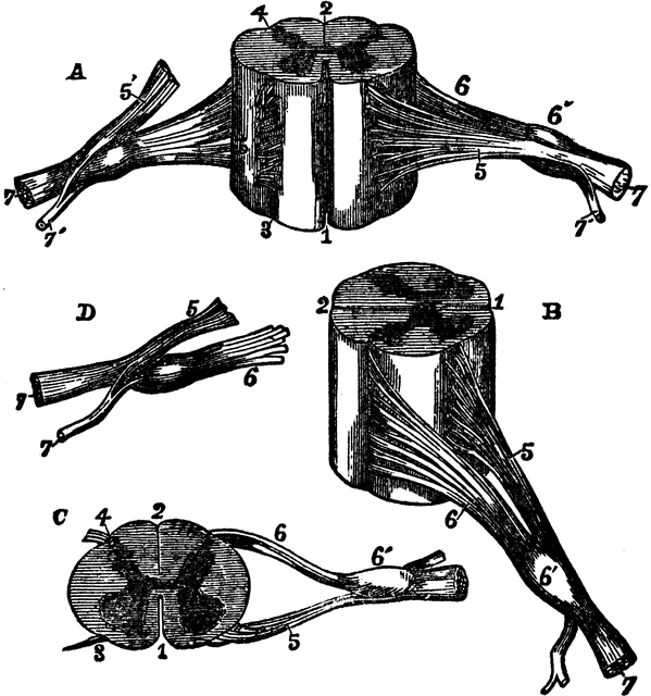
Different views of a portion of the spinal cord from the cervical region, with roots of the nerves slightly enlarged. Labels: In A, the anterior surface of the specimen is shown, the anterior nerve root of its right side being divided: in B, a view of the right side is given; in C, the upper surface is shown; in E, the nerve roots and ganglion are shown from below; 1, the anterior median fissure; 2, posterior median fissure; 3, anterior lateral depression, over which the anterior nerve roots are seen to spread; 4, posterior lateral groove, into which the posterior roots are seen to sink; 5, anterior roots passing the ganglion; 5’, in A, the anterior root divided; 6, the posterior roots, the fibers of which pass into the ganglion 6’; 7, the united or compound nerve; 7’, the posterior primary branch, seen in A and D to be derived in part from the anterior and in part from the posterior root.
Galleries
Human Central Nervous SystemSource
Miller, F. E.; Hunt, H. L.; McCormick, F.J.; Burr, B.; & King, M.L. Domestic Medical Practice: A Household Adviser in the Treatment of Diseases, Arranged for Family Use (Chicago: Domestic Medical Society, 1919) 56
Downloads
2249×2400, 999.5 KiB
959×1024, 151.1 KiB
599×640, 78.3 KiB
299×320, 25.6 KiB