Development of the Primary Optic Vesicle in a ChickClipart ETC: An online service of Florida's Educational Technology Clearinghouse

Development of the Primary Optic Vesicle in a Chick

Development of the Primary Optic Vesicle in a ChickDevelopment of the Primary Optic Vesicle in a ChickDevelopment of the Primary Optic Vesicle in a Chick

To use any of the clipart images above (including the thumbnail image in the top left corner), just click and drag the picture to your desktop. You may also control-click (Mac) or right-click (Windows) and choose "Save file to disk" from the pop-up menu. Click here for help downloading and using clipart files.

Download TIFF* File

* A TIFF file is a high-resolution (240 dpi) format. TIFF files should be used only if you are printing the clipart. Do NOT use TIFF files for clipart that will displayed on a computer screen. Click here for help downloading and using TIFF files.

File Name: 51314_chick
Description: 
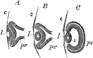
Longitudinal section of the primary optic vesicle in the chick. Labels: A, from an embryo of 65 hours; B, a few hours later; C, of the fourth day; c, the corneous layer or epidermis, presenting in A the open depression from the lends , which is closed in B and C; l, the lens follicle and lens; pr, the primary optic vesicle; in A and B, the pedicle is shown; in C, the section being to the side of the pedicle, the latter is not shown; v, the secondary ocular vesicle and vitreous humour.
Source: Baker, W. Morrant & Harris, Vincent Dormer Kirkes' Hand-book of Physiology, 13th ed. (Philadelphia: P. Blakiston's Son & Co., 1892) 836
Keywords: "optic vesicle", eye

Copyright: 2009, Florida Center for Instructional Technology. See license.

Main MenuSite MapSearchLicenseClipart Help


Clipart ETC is a part of the Educational Technology Clearinghouse and is funded by various grants. Produced by the Florida Center for Instructional Technology, College of Education, University of South Florida. Email the project manager.


Clipart ETCEducational Technology Clearinghouse