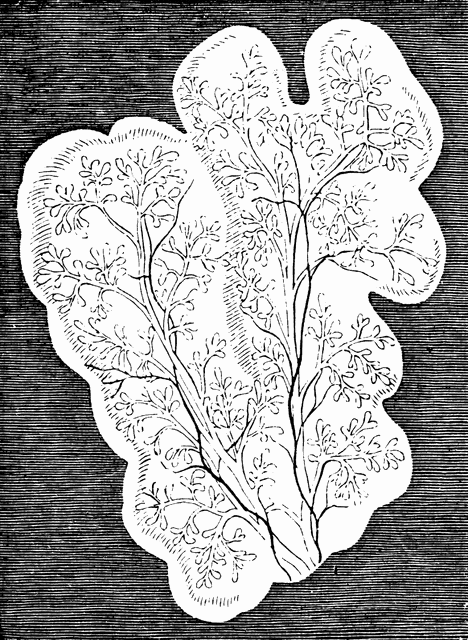
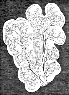
Lobules of Parotid of a Sheep
| View Cart ⇗ | Info

In mammalia, each salivary gland first appears as a simple canal with bud-like processes, lying in a gelatinous nidus or blastema, and communicating with the cavity of the mouth. As the development of the gland advances, the canal becomes more and more ramified, increasing at the expense of the blastema in which it is still enclosed. the branches or salivary ducts constitute an independent system of closed tubes. Shown are lobules of the parotid, with salivary ducts, in the embryo of the sheep at a more advanced stage.
Galleries
Mammal Anatomy: Internal OrgansSource
Baker, W. Morrant & Harris, Vincent Dormer Kirkes' Hand-book of Physiology, 13th ed. (Philadelphia: P. Blakiston's Son & Co., 1892) 841
Downloads
1758×2400, 1.6 MiB
750×1024, 232.4 KiB
468×640, 114.7 KiB
234×320, 37.6 KiB