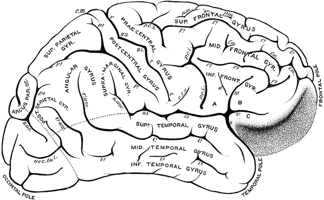
Gyri and Sulci on the Brain

Gyri and sulci, on the outer surface of the cerebral hemisphere. Labels: f1, sulcus frontalis superior; f2, sulcus frontalis inferior; f.m., sulcus frontalis medius; p.m., sulcus paramedialis; A, pars basilaris; B, pars triangularis; C, pars orbitalis; S, Sylvian fissure; s1, anterior horizontal limb (Sylvian fissure); s2, posterior horizontal limb (Sylvian fissure); p.c.i., inferior praecentral sulcus; p.c.s., superior praecentral sulcus; r, fissure of Rolando; g.s., superior genu; g.i., inferior genu; d, sulcus diagonalis; t1, superior temporal sulcus (parallel); t2, inferior temporal sulcus; p1, inferior postcentral sulcus; p2. superior postcentral sulcus; p3, ramus horizontalis; p4, ramus occipitalis; s.o.t., sulcus occipitalis transversus; c.m., callosomarginal sulcus; c.t.r., inferior transverse furrow.
Galleries
Human Central Nervous SystemSource
Cunningham, D.J. Textbook of Anatomy (New York, NY: William Wood and Co., 1903)
Downloads
2400×1478, 683.5 KiB
1024×630, 95.1 KiB
640×394, 48.4 KiB
320×197, 17.4 KiB