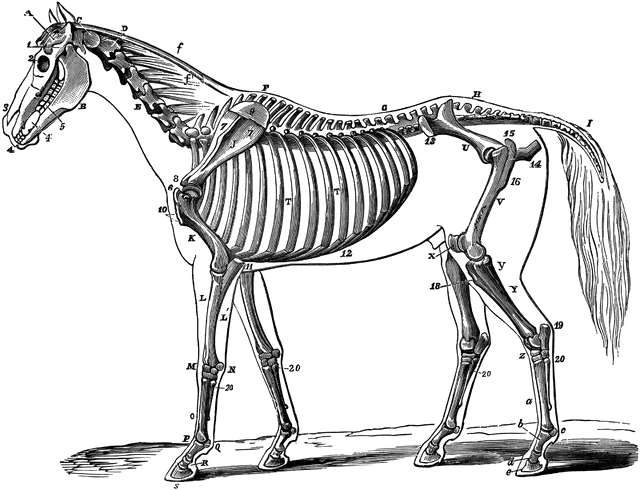
Skeleton of a Horse

The skeleton of a horse. Showing its relation to the contour of the animal, viewed laterally. Labels: A, temporal fossa; B, inferior maxilla; C, atlas; D, dentata; E, cervical vertebrae; F, dorsal vertebrae; G, lumbar vertebrae; H, sacral vertebrae; I, coccygeal vertebrae; J, scapula; K, humerus; L, radius; L, ulna; M, carpus; N, trapezium; O, metacarpus; P, b. os suffraginis; Q, c. sesamoids; R, d. os coronae; S, e. os pedis; T, ribs; U., ilum; V, femur; X, patella; Y, tibia; y., Fibula; Z, tarsus; a, metatarsus; f, ligamentum nuchae, funicular portion; f’ lamellar portion; 1, zygoma; 2, orbital fossa; 3, nasal peak; 4, incisor teeth; 4’, canine teeth; 5, molar teeth; 6, external humeral trochanter; 7, scapular fossae; 8, coracoid apophysis; 9, cartilage of prolongation; 10, deltoid ridge, and external tuberosity; 11, olecranon; 12, costal cartilage; 13, anterior iliac spine; 14, ischium; 15, trochanter major; 16, trochanter minor; 18, anterior tibial tuberosity; 19, calcaneum; 20, small metacarpal and metatarsal or splint bones.
Keywords
horse skeletonGalleries
Mammal Anatomy: SkeletonSource
Vaughn, I. Strangeway's Veterinary Anatomy (Toronto, CAN: J. A. Carveth & Co., Ltd., 1904)
Downloads
2400×1839, 1.3 MiB
1024×784, 168.6 KiB
640×490, 75.6 KiB
320×245, 23.6 KiB