Skull of a Chick Below
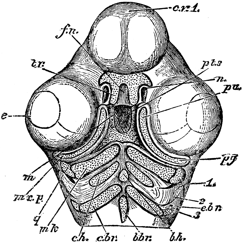
“Skull of a chick, but seen from below. cv1, anterior cerebral vesicle; e, eye; m, mouth; pts, pituitary space; fn, fronto-nasal plate; tr, ends of the trabeculae, free again after their union and bent strongly from the original axis of the trabeculae; n, external nostril; mxp, subocular bar of cartilage, or pterygo-palatine rod, to form pa, palatine, and pg, pterygoid bone, and other parts of the upper jaw, as the maxillary, jugal and quadrato-jugal; q, quadrate cartilage, same as seen in fig 64; mk, meckelian cartilage, to form lower jaw; these parts are in the first post-oral visceral arch; ch, cerato-hyal, and bh, basihyal, of second postoral arch; cbr, cerato-branchial, ebr-branchial, bbr, basi-branchial, of third post-oral arch; the parts of the second and third arch all going into the hyoid bone. 1, 2, 3, 1st, 2d, 3d, visceral clefts, whereof the 1st is to be modified into the ear-passages, and the others are to be obliterated.” Elliot Coues, 1884
Keywords
birds, ornithology, bird anatomy, chick, bird bones, bird skull, North American birds, internal parts of birds, bird jawGalleries
Bird AnatomySource
Elliot Coues Key to North American Birds (Boston, MA: Estes and Lauriat, 1884)
Downloads
2400×2403, 1.3 MiB
1022×1024, 212.4 KiB
639×640, 109.4 KiB
319×320, 37.0 KiB