The Skull of a Chick Stage Three
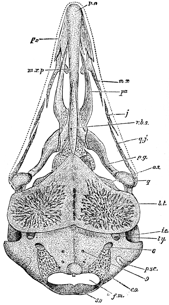
“Skull of chick, third stage, viewed from below, x6 & 2/3 diameters. pn, prenasal cartilage, running behind into the septum nasi; on each side of it the premaxillary, px, of which the (inner) palatal and (outer) dentary processes are seen (the upper nasal process hidden); mx, the maxillary, developing inner process, the maxillo-palatine, mxp; pa, the palatal, well-formed, articulating behind with rbs, the sphenoidal rostrum, its thickened under border, the parasphenoid; this will bear the vomer at its end when that bone is developed; j, jugal, joining mx and qj, the quadrato-jugal, joining j and q, the quadrate; mx to q, the jugal bar or zygoma; pg, the pterygoid, making with pa the pterygo-palatine bar, joining q and px; bt, the basitemporal, great mat of bone from ear to ear, underflooring the skull proper, as rbs, a similar formation, does further forward; ic, outer end of carotid canal, to run between the bt plate and true floor of skull, and enter brain cavity at original site of pituitary fossa; ty, tympanic cavity - external opening of ear; as, alisphenoid, bounding much of brain-box anteriorly, and orbital cavity posteriorly; psc, posterior semicircular canal of ear, in opisthotic bone, which will unite with the spreading eo, exoccipital, which will reach the cobdyle shown in the middle line, above the foramen magnum, fm, completed above by so, supra-occipital; 8, foramen lacerum posterius, exit of pneumogastric, glosso-pharyngeal and spinall accessory nerve; 9, exit of hypoglossal nerve, in basi-occipital.” Elliot Coues, 1884
Keywords
birds, chicks, ornithology, bird anatomy, bird bones, North American birds, chick skull, chick brain, chick bones, bird cartilageGalleries
Bird AnatomySource
Elliot Coues Key to North American Birds (Boston, MA: Estes and Lauriat, 1884)
Downloads
1329×2400, 1.1 MiB
567×1024, 159.3 KiB
354×640, 75.6 KiB
177×320, 21.1 KiB