The Human Sensory Systems: Sight ClipArt gallery offers 189 illustrations related to human vision.
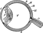
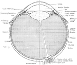
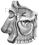
Human Eye
1, the sclerotic thicker behind than in front; 2, the cornea; 3, the choriod; 6, the iris; 7, the pupil;…
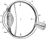
Human Eye
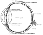
"Diagrammatic horizontal section of the eye of man. c, cornea; ch. choroid (dotted); C. P, ciliary processes;…
Human Eye
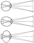
Diagrams of how an image is displayed with a normal eye (top image), myopic or nearsighted eye (middle…

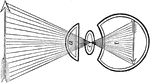
Lens of the Eye
Lens of the eye. The rays of light are brought nearer together by the lenses of the eye, just as they…
Lens of the eye
"Diagram showing the Change in the Lens during Accomadation. On the right the lens is arranged for distant…
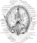
Median Vertical Anteroposterior Section of Eye
"Human Eye, in Median Vertical Anteroposterior Section. (Ciliary processes shown, through not all lying…
Eye, Muscles of
The muscles of the eyeball, which are 6 in number (4 recti and 2 oblique), which are inserted into the…

Muscle of the Eye
Side view of the muscles of the eye in their natural positions. Labels: a,b,c,d, the four straight muscles.…
Muscles of the Eye
The muscles of the right eye. Labels: A, superior straight; B, superior oblique passing through a pulley,…

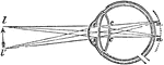
Eye Focusing on Object
"Showing how the image of an object which is seen is formed on the retina of the eye." —Croft 1917
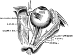
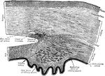
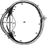
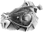
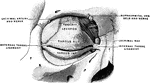
Outer Surface of the Middle Eye
Outer surface of the middle or vascular coat of the eyeball showing the the ciliary gangliated plexus…
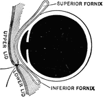
Sagittal Section of the Eye
Sagittal section of the eye, showing superior and inferior fornices of the conjuctiva.
Sagittal Section Through the Eye
The upper half of a sagittal section through the front of the eyeball.
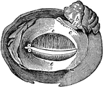
Section of the Eye
Section of the eye magnified, showing the ciliary processes, the pigmentum nigrum, the retina, and the…
Section of the Eye
The vitreous humor and crystalline lens of the eyeball magnified, with the stains of the pigmentum nigrum…
Section of the Eye
Section of the eye magnified, showing the crystalline lens in its proper situation, between the aqueous…
Section of the Eye
Section of the eye, showing the relations of the cornea, sclera, and iris, together with the Ciliary…
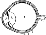
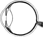
The Eye
The eye. Labels: a, sclerotica; e, cornea; b, choroid; d, optic nerve; f, aqueous humor; g g , iris;…
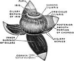
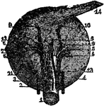
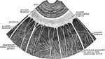
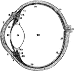
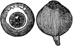
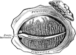
Vascular Coat of the Eye
The middle or vascular coat of the eyeball exposed from without. Left eye, seen obliquely from above…
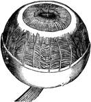
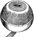
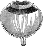
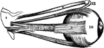
Eyeball
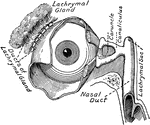
"The Relative Position of the Lachrymal Apparatus, the Eyeball, and the Eyelids. A, lachrymal canals,…
Eyeball
Section through the closed left eye. 1. Lifting muscle 2. Upper Straight Muscle 3. Optic Nerve 4. Fatty…
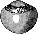
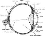
Eyeball
"The most essential parts of human vision are contained in the eyeball, a nearly spherical body, about…
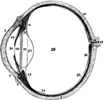
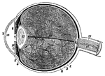
Left Eyeball in Horizontal Section
The left eyeball in horizontal section from before back. Labels: 1, sclerotic; 2, junction of sclerotic…
Section of the Eyeball
Section of the eyeball. Labels: Con, conjunctiva; C, cornea; A, aqueous humor; I, iris; L, crystalline…
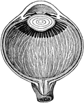
Eyeball
Horizontal section of the eyeball, showing the suspensory ligament of the lens, the aqueous and vitreous…
The Eyeball in Horizontal Section
The left eyeball in horizontal section from before back. Labels: 1, sclerotic; 2, junction of sclerotic…
Section of Left Eyeball
The left eyeball in horizontal section from before back. Labels: 1, sclerotic; 2, junction of sclerotic…
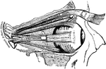
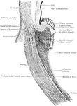
The Eyeball with its Muscle
The eyeball with its muscles attached. The upper muscle not attached to the ball belongs to the upper…
Muscles of the eyeball
"A, attachment of tendon connected with the four recti muscles; B, external rectus,…
Side View of the Eyeball
Side view of the eyeball. Labels: a, the eyeball, and b,b, are the upper and lower sides. Now in order…
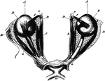
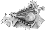
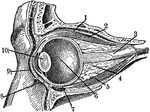
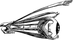
The Eyeballs and Their Muscles
The eyeballs and their muscles as seen when the roof of the orbit has been moved and the fat in the…

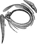
Eyelashes
This diagram shows the eyelids and the tear-glands. The eyelid is a thin fold of skin and muscle that…
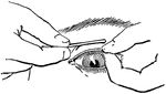
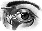
Everted eyelid
"Showing how the upper eyelid may be everted with a pencil or penholder." — Blaisedell, 1904
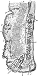
Vertical Section Through Eyelid
Vertical section through the upper eyelid. Labels: a, Skin; b, Orbicularis palpebrarum; b', Marginal…
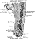
Eyelids, Viewed from Behind
The eyelids separated, and viewed from behind. Labels: a, the lachrymal gland; b, the ducts from the…
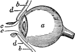
Eyelids, Viewed from the Front
The eyelids viewed from before; a,a, the lachrymal canals; b, the lachrymal sack. The lachrymal sac…
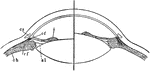
Flattened Eye
"A representation of the manner in which the image is formed in the eye, when the cornea or crystalline…
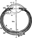
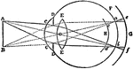
Focusing of the Eye
Diagram to illustrate the mechanism of accommodation (focusing); on the right half of the figure for…
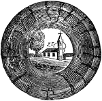
Formation of an Image on the Eyeball
Suppose a person was looking at a church with a tree standing at its side, he would have in each eye…
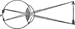
Formation of an Image on the Eyeball
In passing through the crystalline, the rays cross each other, so that those rays which pass from the…
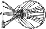
Formation of Circles of Diffusion
An illustration depicting the formation of circles of diffusion. "From point A luminous rays enter the…
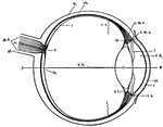
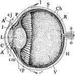
Human Eye
This diagram shows a side view of the right eye of man. a.c., central artery; a.h., aqueous humor; b.,…
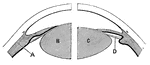
Indistinct Vision
"...where we suppose that the object a, is brought within an inch or two of the eye, and that the rays…
Inversion of Objects by the Eye
"The actual position of the vertical object a, as painted on the retina, is therefore such as is represented…