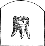
This human anatomy ClipArt gallery offers 82 illustrations of human teeth and jaws. This includes external and internal views focusing on human adult and juvenile teeth. Views of teeth within the jaw bones are also included.

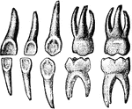
The permanent teeth of the right side. The numbers show at what age they appear. Labels: a, incisors; b, canines; c, premolars; d, molars.

Permanent Teeth

The permanent teeth of the right side. The numbers show at what age they appear. Labels: a, incisors;…

Permanent teeth, right side.

Permanent Teeth

Permanent teeth, right side.

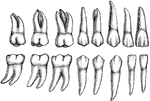
The permanent teeth of the right side, outer or labial aspect. The upper row shows the upper teeth, the lower row the lower teeth. The wide vertical labial ridge is distinct on the upper canine and premolar teeth.

Permanent Teeth

The permanent teeth of the right side, outer or labial aspect. The upper row shows the upper teeth,…

The permanent teeth of the right side, inner of lingual aspect. The upper row shows the upper teeth, the lower row the lower teeth. The cingulum is distinct on the upper incisors and both canines, the lingual cusp on the upper lateral incisor and the upper canine.

Permanent Teeth

The permanent teeth of the right side, inner of lingual aspect. The upper row shows the upper teeth,…

"A Tooth is one of the hard bodies of the mouth, attached to the skeleton, but not forming part of it and developed from the dermis or true skin. True teeth consist of one, two, or more tissues differing in their chemical composition and in their microscopical appearances. Dentine, which forms the body of the tooth, and 'cement,' which forms its outer crust, are always present, the third tissue, the 'enamel,' when present, being situated between the dentine and cement. The incisors, or cutting teeth, are situated in front. In men there are two of these incisors in each side of each jaw. The permanent incisors, molars, and premolars are preceded by a set of deciduous or milk teeth, which are lost before maturity, and replaced by the permanent ones. The canines come next to the incisors. In man there is one canine tooth in each half-jaw. The premolars (known also as bicuspids and false molars) come next in order to the canines. In man there are two premolars in each half-jaw. The true molars (or multicuspids) are placed most posteriorly. In man there are three molars in each half-jaw, the posterior one being termed the wisdom tooth. The figures [in the illustration] refer to years after birth."—(Charles Leonard-Stuart, 1911)

Second Teeth

"A Tooth is one of the hard bodies of the mouth, attached to the skeleton, but not forming part of it…

Structure of the teeth. A tooth consists of 3 structures, the dentine (2), or ivory, the proper dental substance, forming the larger portion; the enamel or adamant (3), a layer covering the crown; and the cement (1), a thin layer on the surface of the fang. A central chamber, the pulp-cavity, occupies the interior, and contains the pulp, a soft, vascular, and sensitive organ, surrounded by a layer of cells, the odonto-blasts, or dentine-forming cells.

Structure of the Teeth

Structure of the teeth. A tooth consists of 3 structures, the dentine (2), or ivory, the proper dental…

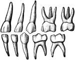
The temporary (baby) teeth. On the left are the teeth of the left side of the mouth viewed from the outside. On the right are teeth of the right side of the mouth viewed from the inside of the mouth.

Temporary Teeth

The temporary (baby) teeth. On the left are the teeth of the left side of the mouth viewed from the…
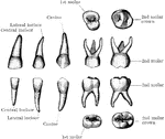
Labial view of temporary teeth of the left side.

Temporary Teeth

Labial view of temporary teeth of the left side.

Lingual view of temporary teeth.

Lingual View of Temporary Teeth

Lingual view of temporary teeth.

The temporary teeth of the left side. The masticating surfaces of the tow upper molars are shown above. In the second row the upper teeth are viewed from the outer or labial side. In the third row the lower teeth are shown in a similar manner; and below are the masticating surfaces of the two lower molars. In the specimen from which the first upper molar was drawn the two outer or buccal cusps were not distinctly separated, as is often the case.

Temporary Teeth

The temporary teeth of the left side. The masticating surfaces of the tow upper molars are shown above.…
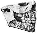
"<em>Temporary Teeth:</em> <em>A</em>, central incisors; <em>B</em>, lateral incisors; <em>C</em>, canines; <em>D</em>, anterior molars; <em>E</em>, posterior molars, <em>F</em>, central incisors; <em>H</em>, lateral incisors; <em>K</em>, canines; <em>L</em>, first bicuspids; <em>M</em>second bicuspids; <em>N</em>, first molars." &mdash; Blaisedell, 1904

Temporary and permanent teeth

"Temporary Teeth: A, central incisors; B, lateral incisors; C, canines;…

Characteristics of the teeth. Teach tooth consists of a crown or body, projecting above the gum; root or fang, embedded within the alveolus; and the neck, the constricted portion between the grown and the fang. There are 4 classes of teeth: incisors (1), canines (2), premolars or bicuspids (3), and molars (4).

The Teeth

Characteristics of the teeth. Teach tooth consists of a crown or body, projecting above the gum; root…
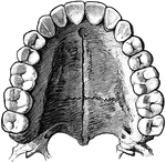
The upper permanent teeth.

Upper Permanent Teeth

The upper permanent teeth.

A small bone attached to the jaw for chewing.

Tooth

A small bone attached to the jaw for chewing.

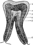
Vertical section of a tooth. 1: Enamel; 2: Dentine; 3: Pulp; 4: Blood-vessel; 5: Nerve; 6: Fibrous cement.

Tooth

Vertical section of a tooth. 1: Enamel; 2: Dentine; 3: Pulp; 4: Blood-vessel; 5: Nerve; 6: Fibrous cement.

Diagram to illustrate the development of a tooth. I. Shows the downgrowth of the dental lamina D.L. from the surface epithelium E and the beginning of the enamel germ E.G. II. Shows the further growth of the enamel germ and its invagination. III. The enamel germ is more invaginated, and its inner layer of cells becomes columnar. A, the dental lamina, grows thinner, but near its posterior or lingual edge there is an enlargement R.G which is the reserve germ for a permanent tooth. The superficial cells of the dentine papilla P are becoming columnar. IV. The inner columnar cells of the enamel germ ( called enamel cells) A have formed a cap of enamel EN, inside which the superficial cells of the papilla, the odontoblasts O, have formed a layer of dentine D. V. Shows a more advances stage still. The deposit of dentine is extending downwards, and enclosing the papilla to form the future pulp, in which a vessel V is seen. Labels: A, inner layer; B, outer layer; C, remains of intermediate cells; D, dentine; D.L, dental lamina; E, epithelium; E.G, enamel germ; EN, enamel; F, dental furrow; L.D, labio-dental furrow; M, connective tissue cells; O, odontoblasts; P, dentine papilla; R.G, reserve germ; V, blood vessels.

Development of a Tooth

Diagram to illustrate the development of a tooth. I. Shows the downgrowth of the dental lamina D.L.…
A tooth is generally described as possessing a crown, neck, and root. Side view of a tooth.

Human Tooth

A tooth is generally described as possessing a crown, neck, and root. Side view of a tooth.
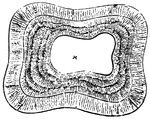
A tooth is generally described as possessing a crown, neck, and root. Top view of a tooth.; 1. Central Nippers, worn to a plain surface.; 2. next pair still sowing a slight remnant of the cavity.; 3. Corner nippers, showing the mark plainly enough.; 4. Tushes, more worn down that in the lower jaw of the six-year-old mouth.

Human Tooth

A tooth is generally described as possessing a crown, neck, and root. Top view of a tooth.; 1. Central…
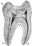
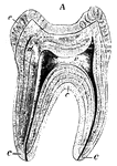
A sectional view of a human molar. The roots, or fangs, are shown covered by a layer of bone called "cement", which is represented by letter c. The center of the tooth is the vascular pulp-cavity v, d is ivory or dentine, and e is the hardest tissue of the body - enamel.

Human Tooth

A sectional view of a human molar. The roots, or fangs, are shown covered by a layer of bone called…
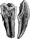
"<em>A</em>, enamel; <em>B</em>, pulp cavity; <em>C</em> dentine; <em>D</em>, cement." &mdash; Blaisedell, 1904

Longitudinal section of a tooth

"A, enamel; B, pulp cavity; C dentine; D, cement." — Blaisedell,…

The internal view of a tooth cut through from the top or crown to the tips of the root. Labels: 1, enamel; 2, dentine; 3, pulp

The Structure of a Tooth

The internal view of a tooth cut through from the top or crown to the tips of the root. Labels: 1, enamel;…

Vertical section of a tooth in situ. Labels: c, pulp cavity; 1, enamel with radial and concentric markings; 2, dentin with tubules and incremental lines; 3, cementum or crusta petrosa; 4, pericemental membrane; 5, bone of mandible.

Vertical Section of a Tooth

Vertical section of a tooth in situ. Labels: c, pulp cavity; 1, enamel with radial and concentric markings;…