This human anatomy ClipArt gallery offers 82 illustrations of the human excretory system, including views of the systems involved in excreting waste that are not already included in the respiratory and digestive systems. Included here are the urinary tract, renal system (e.g., kidneys), and excretory glands of the skin (e.g., sebaceous and sweat glands).
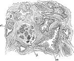
Epithelial Elements of a Malpighian Capsule
Epithelial elements of a Malpighian capsule and tuft, with the commencement of a urinary tubule showing…
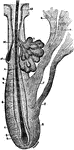
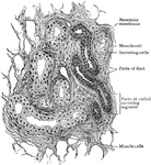
Section of the Prostate Gland
Section of a small portion of the prostate. Labels: a, gland duct cut across obliquely; b, gland structure;…
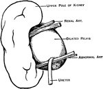
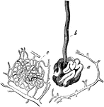
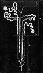
Abnormal Renal Artery
An abnormal renal artery causing kinking at the ureteropelvic junction, and hydronephrosis.
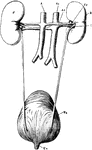
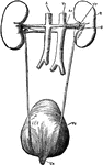
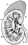
Renal Organs
"The renal organs, viewed from behind. R, right kidney; A, aorta; Ar, right renal artery; Vc, inferior…
The Renal Organs Viewed from Behind
The renal organs viewed from behind. labels: R, right kidney; A, aorta; Ar, right erenal artery; Vc,…
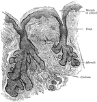
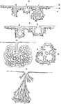
Secreting Gland
Secreting glands may be classified according to certain types. Shown are plans of extension of secreting…
Section of Human Kidney
This illustration shows a section of a human kidney (A, Cortical substance; B, Pyramids; C, Hilum; D,…
Sweat Gland
Coiled end of a sweat gland. Labels: a, the coiled end; b, the duct; c, network of capillaries, inside…
A Sweat Gland
A sweat gland. Labels: d, horny layer of cuticle; c, Malpighian layer; b, dermis. The coils of the gland…
Sweat Gland
A sweat gland. Labels: a, horny layer of cuticle; c, Malpighian layer; b, dermis. The coils of the gland…
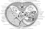
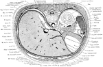
Cross Section of the Trunk through Costal Arch
Section passes through the lower portion of the costal arch, and through the hilus of the two kidneys.
Cross Section of the Trunk through Third Lumbar Vertebra
Section through the third lumbar vertebra and the inferior poles of the kidneys cutting the loop of…
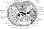
Cross Section of the Trunk through Upper Kidney
Section through the upper pole of the left kidney at the level of the tip of the xiphoid process.
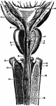
Entrance of Ureter into the Bladder
Diagram showing the method of entrance of the ureter into the bladder.
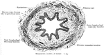
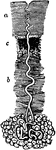
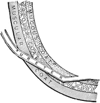
Transverse Section of Ureter
In human anatomy, the ureters are muscular tubes that propel urine from the kidneys to the urinary bladder.
The Male Urethra
The male urethra, which is the urinary canal from the neck of the bladder to the meatus urinarius; in…
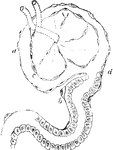
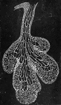
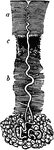
Uriniferous Tubes
A diagram of the sections of uriniferous tubes. A, Cortex limited externally by the capsule; a, subcapsular…