This human anatomy ClipArt gallery offers 32 illustrations of the human endocrine system, which includes the system of organs involved in the production of hormones responsible for regulating metabolism, growth, development, and tissue function that are not included in the other systems. Organs in this category include the thyroid, parathyroid, thymus, adrenal glands, liver, and pancreas.

Diagram of two hepatic lobules of the liver. "The left hand lobule is represented with the intralobular vein cut across; in the right hand one the section takes the course of the intralobular vein. p, interlobular branches of the portal vein; h, intralobular branches of the hepatic veins; s, sublobular vein; c, capillaries of the lobules. The arrows indicate the direction of the course of the blood. The liver cells are only represented in one part of each lobule." — Kimber, 1907.

Hepatic Lobules of the Liver

Diagram of two hepatic lobules of the liver. "The left hand lobule is represented with the intralobular…
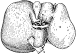
"The Liver seen from Below and Behind. <em>A</em> and </em>B</em>, smaller lobes of the liver; <em>C</em>, portal vein; <em>D</em> hepatic artery; <em>E</em>, inferior vena cava; <em>F</em>, trunk of the left hepatic vein; <em>G</em>, hepatic vein." &mdash; Blaisedell, 1904

Liver

"The Liver seen from Below and Behind. A and B, smaller lobes of the liver; C,…

Under surface of liver. Labels: 1, right lobe; 2, left lobe; 3, 4, 5, smaller lobes; 9, inferior vena cava; 10, gall-bladder; 11, 11, transverse fissure, or "gate of the liver," containing bile duct, hepatic artery, and portal vein.

Under Surface of Liver

Under surface of liver. Labels: 1, right lobe; 2, left lobe; 3, 4, 5, smaller lobes; 9, inferior vena…
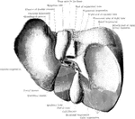
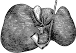
The liver, lower surface.

Liver

The liver, lower surface.

The liver from the below and behind, showing the whole of the visceral surface and the posterior area of the parietal surface. The portal fissure has been slightly opened up to show the vessels passing through it; the other fissures are represented in their natural condition-closed. In this liver, which was hardeded in situ, the impressions of the sacculations of the colon were distinctly visible at the colic impression. The round ligament and the remains of the ductus venosus are hidden in the depths of their fissures.

Liver from Behind

The liver from the below and behind, showing the whole of the visceral surface and the posterior area…
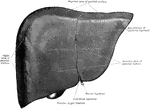
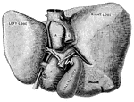
The liver from the front, showing the superior, right, and anterior areas of the parietal surface.
Liver from Front

The liver from the front, showing the superior, right, and anterior areas of the parietal surface.

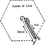
Diagram to illustrate the relationship of blood capillaries, bile capillaries, and liver cells. Labels: L, intralobular vein at center of lobule; P, vessel of interlobular plexus from which a blood capillary extends to L; D, a minute bile duct with which a bile capillary communicates.

Liver Structure

Diagram to illustrate the relationship of blood capillaries, bile capillaries, and liver cells. Labels:…
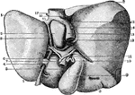
Deformed female liver.

Deformed Liver

Deformed female liver.

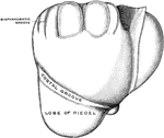
Fissures and Lobes of the liver. There are 5 fissures of the liver, which are situated on the inferior and posterior surfaces and arranged in the form of the letter H. There are 5 lobes of the liver, but 3 are mere lobules of the right lobe, formed by the smaller fissures.

Fissures and Lobes of the Liver

Fissures and Lobes of the liver. There are 5 fissures of the liver, which are situated on the inferior…

Diagram to illustrate the arrangement of the blood vessels (on right) within the lobule of the liver. The first diagram shows the interlobular veins running around the outerside of the lobule, and sending their capillaries into the lobule to join the central radiation to the periphery of the lobule, where they can join the interlobular bile ducts.

Structure of Liver

Diagram to illustrate the arrangement of the blood vessels (on right) within the lobule of the liver.…
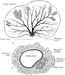
A diagram to illustrate the structure of the liver. A, arrangement of liver lobules around the sublobular branches of the hepatic vein; B, Section of portal canal, showing its contained branches of the portal vein, hepatic artery, and bile duct, surrounded by prolongation of Glisson's capsule.

Structure of the Liver

A diagram to illustrate the structure of the liver. A, arrangement of liver lobules around the sublobular…
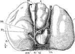
The under surface of the liver. Labels: d, right, and s, left lobe; Vh, hepatic vein; Vp, portal vein; Vc, vena cava inferior; Dch, common bile-duct; Dc, cystic duct; Dh, hepatic duct; Vf, gallbladder.

The Under Surface of the Liver

The under surface of the liver. Labels: d, right, and s, left lobe; Vh, hepatic vein; Vp, portal vein;…

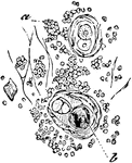
Transverse section of a lobule of an injected infantile thymus gland. Labels: a, capsule of connective tissue surrounding the lobule; b, membrane of the glandular vesicles; c, cavity of the lobule, from which the large blood vessels are seen to extend towards and ramify in the spheroidal masses of the lobule.

Lobule of Thymus Gland

Transverse section of a lobule of an injected infantile thymus gland. Labels: a, capsule of connective…
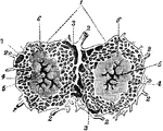
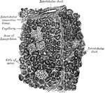
Lobules of the liver (1), which are small, granular-looking bodies, of polygonal shape, and about 1/20 inch in diameter, clustered around the sublobular branches of the hepatic veins, and connected together by connective tissue (3), blood-vessels, ducts (2), and lymphatics. Each lobule consists of a mass of hepatic cells, surrounded by a dense capillary plexus, and contains the minute beginnings of a bile-duct, the so-called biliary capillaries; possessing therefore all the essential constituents of a secreting gland. A lobule contains hepatic cells, lobular veins (3), intralobular veins (4), and plexuses of lymphatics, nerves, and bile-ducts.

Lobules of the Liver

Lobules of the liver (1), which are small, granular-looking bodies, of polygonal shape, and about 1/20…
"The pancreas, partly cut away, so as to show the duct, which collects the pancreatic juice, and empties it into the duodenum." &mdash; Tracy, 1888

Pancreas

"The pancreas, partly cut away, so as to show the duct, which collects the pancreatic juice, and empties…
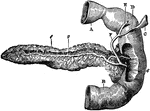
Posterior view of pancreas. Labels: 1, pancreas; 2, pancreatic duct; 6, opening of common duct, formed by union of pancreatic and choledochus ducts, into duodenum; A, pyloric end of stomach; B, duodenum; C, part of gall-bladder; D, cystic duct; E, hepatic duct; F, choledochus duct.

Posterior View of Pancreas

Posterior view of pancreas. Labels: 1, pancreas; 2, pancreatic duct; 6, opening of common duct, formed…

Section of pancreas under low magnification, showing general arrangement of lobules.

Section of Pancreas Showing Arrangement of Lobules

Section of pancreas under low magnification, showing general arrangement of lobules.

Section of human pancreas, showing pancreatic islands.

Section of Pancreas

Section of human pancreas, showing pancreatic islands.

The pancreas, a compound racemose gland about 5.5 inches long and 1.5 inches wide, situated transversely across the posterior wall of the abdomen behind the stomach and in front of the first lumbar vertebra.

The Pancreas

The pancreas, a compound racemose gland about 5.5 inches long and 1.5 inches wide, situated transversely…
The position of the parathyroid glands. Viewed from behind.

Parathyroid Gland

The position of the parathyroid glands. Viewed from behind.

The thymus gland in a full term fetus.

Thymus Gland

The thymus gland in a full term fetus.

Deep surface of the thymus gland, taken from a fetus.

Thymus Gland

Deep surface of the thymus gland, taken from a fetus.

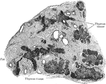
Section of thymus body of man of twenty eight, showing invasion and replacement of thymus tissue by fat.

Thymus Showing Replacement of Thymus Tissue by Fat

Section of thymus body of man of twenty eight, showing invasion and replacement of thymus tissue by…

Elements of the thymus.

Elements of the Thymus

Elements of the thymus.

A lobule of the thymus of a child, as seen under low power. Labels: C, cortex; c, concentric corpuscles within medulla; b, blood vessels; tr, trabeculae.

Lobe of the Thymus

A lobule of the thymus of a child, as seen under low power. Labels: C, cortex; c, concentric corpuscles…

Transverse section of thymus of child, showing general arrangement of lobules.

Transverse Section of Thymus

Transverse section of thymus of child, showing general arrangement of lobules.

A side view of the thyroid and cricoid cartilages.

Thyroid

A side view of the thyroid and cricoid cartilages.

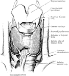
Dissection of the thyroid body and of the parts in immediate relation to it.

Thyroid Body

Dissection of the thyroid body and of the parts in immediate relation to it.

The thyroid gland.

Thyroid Gland

The thyroid gland.

Section through the thyroid gland and the upper borders of the shoulders.

Cross Section Through the Trunk at the Shoulders

Section through the thyroid gland and the upper borders of the shoulders.

Section through the body of the liver at the level of the arch of the seventh rib anteriorly.

Cross Section of the Trunk through the Liver

Section through the body of the liver at the level of the arch of the seventh rib anteriorly.
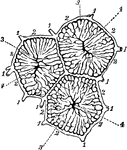
Vessels of the liver. Labels: (1) portal canals, (2) interlobular plexus, (3), lobular veins, (4), intralobular vein.

Vessels of the Liver

Vessels of the liver. Labels: (1) portal canals, (2) interlobular plexus, (3), lobular veins, (4), intralobular…