Sponges
The Sponges ClipArt gallery includes 48 illustrations of sponges. Sponges, also called poriferans, are in the phylum porifera and are all sessile animals that live and feed attached to the bottom of the sea.
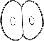
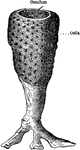
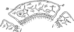
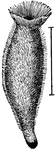
A. Primordialis
"Simple sponge (Ascetta primordialis). Note the vase-like form, the apical osculum, the inhalant pores…
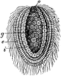
Ascon Sponge
Diagram showing the Ascon type of sponge. ec., ectoderm; r.c., radiating canals; i.p., internal pores;…
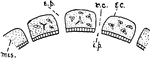
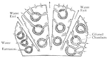
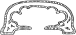
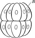
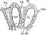
Sponge Canal System
"Diagram showing types of canal system. --After Korschelt and Heider. The flagellate regions are dark…
Chalk Sponge Gastrula
"Gastrula of a Chalk-sponge (Olynthus). B, longitudinal section through the axis: g, primitive intestine;…
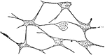
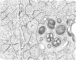
Connective Tissue
"Connective tissue of sponge." — Encyclopedia BSponge Diver Counting Cardsritannica, 1893
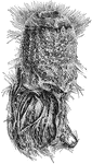
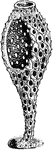
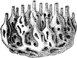
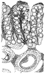
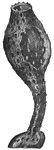
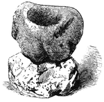
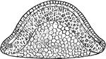
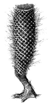
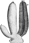
Holtenia Carpenteri
A modern sponge with silicious skeletal structure, and basal glass fibers for fixation (Holtenia…
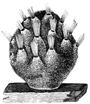
O. Lobularis
"Diagrammatic representation of development of Oscarella lobularis. Bl., Free-swimming blastula with…
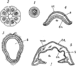
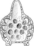
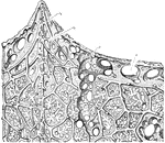
Sponge Section
"Section of a sponge. Showing inhalant canals, flagellate chambers, a gastrula forming in the mesogloea,…
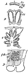
Simple Sponge Development
"Diagrams to illustrate the development of one of the simpler types of sponge: I, the egg; 2, section…
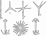
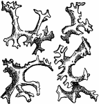
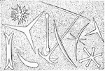
Sponge Spicules
"Sponge spicules. 1, Monaxon; 2, triod; 3, triaxon; 4, tetraxon; 5, anchor; 6, polyaxon; 7, a kind of…
Sponge Spicules
Spicules may be made of silica or calcium carbonate, and vary in shape from simple rods to three-dimensional…
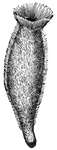
Sponge
In this simple sponge, water enters minute holes in the sides and passes out of the opening at the top…
Sponge
"Diagram of simple type of sponge. c, cloaca; ch, chambers, lined with flagellate entoderm; e.p., external…
Sponge
The young sponge, a many celled animal, that begins its life as a single-cell, the egg. A section of…
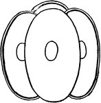
Sponge
Calcolynthus primigenius, a simple sponge with the wall removed to show the inside. It is hollow, attached…
Sponge Develoment
The sponge, a many celled animal, begins its life as a single-cell, the egg. First division in into…
Sponge Develoment
The sponge, a many celled animal, begins its life as a single-cell, the egg. Second division into the…
Sponge Develoment
The sponge, a many celled animal, begins its life as a single-cell, the egg. Fourth division into a…
Sponge Develoment
The sponge, a many celled animal, begins its life as a single-cell, the egg. The continued division…
Sponge Develoment
The sponge, a many celled animal, that begins its life as a single-cell, the egg. The continued division…
Sponge Develoment
The sponge, a many celled animal, begins its life as a single-cell, the egg. After division of cells…
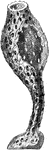
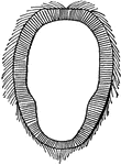
A Cretaceous Sponge
A Cretaceous sponge with silicious skeleton retaining its form. (Ventriculites simplex).
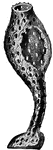
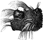
Glass Sponge
A beautiful Glass Sponge called the "Venus Flower Basket". It grows in the deep sea near the Philippine…
Living sponge, magnified
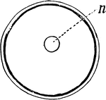
"A glace at a piece of common sponge will show that its surface is everywhere perforated with an infinite…
Lower Section of Sponge
An illustration of the lower portion of a sponge. O, OS, and M are illustrations of sponge eggs magnified…
Skeleton of Sponge
In zoology a skeleton is any fairly rigid structure of an animal, irrespective of whether it has joints…
Upper Section of Sponge
An illustration of the upper portion of a sponge: p, Pore; s, Subdermal cavity; c1, chief fiver of the…
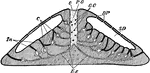
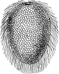
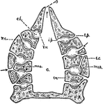
Spongilla
"Vertical section of a fresh-water sponge (Spongilla), showing the arrangement of the canal-system.…

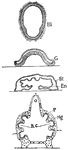
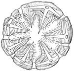
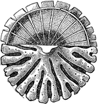
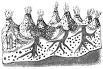
Sycon Gelatinosum
"Transverse section through the wall of a cylinder (parallel with the course of the canals), showing…
Sycon Sponge
Diagram showing the Sycon type of sponge. ec., ectoderm; r.c., radiating canals; i.p., internal pores;…