This human anatomy ClipArt gallery offers 265 illustrations of the central nervous system, including external and dissected views of the brain and spinal cord.
Ventral and Dorsal Views of the Spinal Cord
Diagram of the spinal cord and its fissure. The figure to the left is a ventral aspect and the figure…
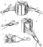
Views of a Spinal Cord
Different views of a portion of the spinal cord from the cervical region, with the roots of the nerves.…
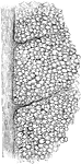
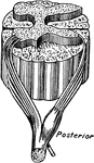
White Matter of Spinal Cord
Transverse section through the white matter of the cord, as seen through the microscope.
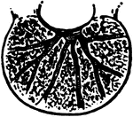
Spinal Marrow
The spinal marrow. Labels: a, Spinal marrow. b, Fibers of sensation. c, Fibers of motion. e, Nerve.
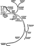
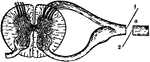
Course of the Spinal Marrow
View of the course of the front columns of the spinal marrow terminating in the hemispheric ganglions…
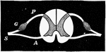
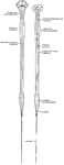
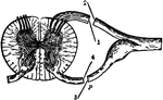
Spinal Nerve
A spinal nerve. Labels: P, posterior root of a spinal nerve; G, ganglion; A, anterior root; S, spinal…
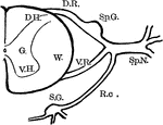
Spinal Nerve Roots
Diagram showing anatomy of the spinal nerve roots and adjacent parts. Labels: G., gray matter of the…
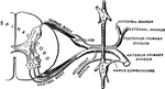
Spinal Nerves
Illustrating the functions of the spinal nerves. Divided at a. -- Irritated at 1: pain. Irritated at…
Spinal Nerves
Diagrammatic representation of the roots and ganglia of the spinal nerves, showing their position in…
Roots of Spinal Nerves
Illustrating the functions of the roots of the spinal nerves. Labels: a, ventral root; p, dorsal root.…
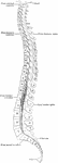
Spine
"The spine, sawn in two lengthwise, showing the spinal canal and the holes between the vertebrae, where…
Spine
Diagram on frozen section, showing relations of bodies and spines of vertebrae to levels at which spinal…
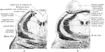
Section Through Tegmentum
Two sections through the tegmentum of the pons at its upper part, close to the mesencephalon. A is at…
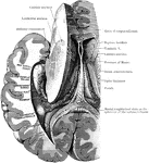
Dissection to Show Ventricle and Fornix
Dissection, to show the fornix and the posterior and descending cornua of the lateral ventricle of the…
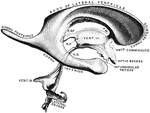
Dissection to Show Ventricle
Dissection, to show the posterior and descending cornua of the lateral ventricle. Labels: B.G., Giacomini's…
Ventricles of the Brain
Cast of the ventricles of the brain. Labels" R.SP., recessus suprapinealis; R.P., recessus pinealis…
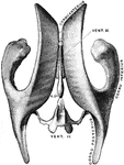
Ventricles of the Brain
Drawing taken from a cast of the ventricular system of the brain, as seen from above. Vent. III, Third…
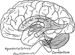
Relations of the Ventricles to the Surface of the Brain
Scheme showing relations of the ventricles to the surface of the brain.
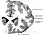
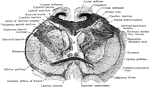
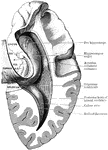
Section Through Lateral Ventricles
Coronal section through the frontal lobes and the anterior horns of the lateral ventricles.