This ClipArt gallery offers 142 illustrations of the anatomy of fish, including organ diagrams, skeletal diagrams, fish eggs, and more for numerous species.
Abdominal
"Pertaining to the abdomen or belly; situated in or on the abdomen: as abdominal ventral fins."-Wright,
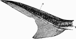
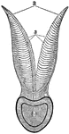
Acipenser
"Heterocercal tail of Acipenser. a, fulcra; b, osseous bucklers." — Encyclopedia Britannica, 1893
The Alimentary Canal of the Herring
There are two predominant forms of stomach in fish- one is like a bent tube (siphonal), and the other…

The Alimentary Canal of the Swordfish
In some species of fish the small intestine is in two to eight coils. The large intestines are short…
A Diagram of the Circulation of the Blood Through the Branchial Leaflets in a Fish
A diagram of the circulation of the blood through the branchial leaflets in a fish. Labels: 1. A section…
Brachiostoma
"Anterior end of body of Branchiostoma. d, chorda dorsalis; e, spinal cord; f, cartilaginous rods; g,…

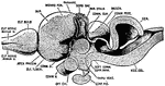
Brain of a Fish
The brain of fish are small, it does not fill the whole cranial cavity, there being found within it…
Section of a Branchial Arch of a Cod
In breathing, the mouth and gills of a fish open alternately; the water entering the mouth escapes by…
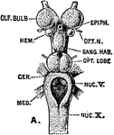
Carcharias Brain
"Brain of Carcharias. ae, nervus acousticus; b, corpus restiforme; c, cerebellum; d, lobus opticus;…
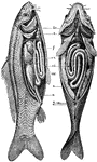
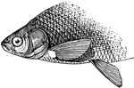
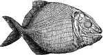
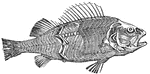
Anatomy of the Carp
br: the branchiae, or gill-openings c: the heart f: the liver vn: swimming bladders ci: intestinal canal…
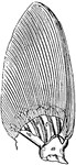
Cycloid Scale of the Carp
Fishes are sometimes classed, in accordance with the structure of their scale, into Ctenoid, Ganoid,…
Ceratodus Lung
"Lung of Ceratodus, opened in its lower half to show its cellular pouches. a, right half; b, left half;…
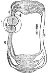
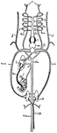
Circulation of a Fish
A diagram of the circulation of a fish. Labels: 1, The pericardium. 2, The single auricle. 3, The single…

Circulation of a Fish
Diagram of the circulation of the fish. Labels: a, branchial circulation; b, somatic circulation; c,…
Cod Skull
"The skull of a cod. b, branchiostegal rays born on c.h., the ceratohyal bone; d, dentary portion of…
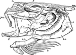
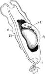
Cuttlefish Organs
"Central organs of the circulaion, gills, and renal organs of Sepia officinalis. a, aorta; v, vena cava;…
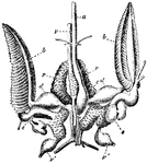
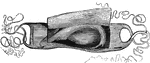
Bone of the cuttle-fish
"An internal support of a calcareous nature, and formed in laminae; this is the well-known cuttlefish…


Ganoid Scales of Dipterus
Fishes are sometimes classed, in accordance with the structure of their scale, into Ctenoid, Ganoid,…
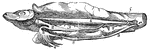
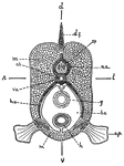
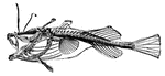
Dissected Fish
"Dissected fish. a, air bladder; b, urinary bladder; b, urinary bladder; br, brain; c, spinal cord;…
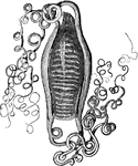
Dogfish Embryo
"Embryo dogfish in egg-case ("mermaid's purse") which has been cut open to show contents. e.g., "External"…
Eel Development
"Development of eel. Change from Leptocephalus shape (I.) to "Elver" shape (V.)." -Thomson, 1916
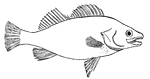
Fish
Outline of a fish, showing the "paired" and "median" fins. (p) one of the pectoral fins; (v) one of…
Fish
This is a diagram of the cross section of a fish, showing the bilateral symmetry of the parts: dv, dorsoventral…
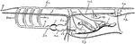
Fish Circulation
"Diagram of the principal vessels in the circulation of a fish, ventral view. a, aorta; au., auricle;…
Fish Circulation Vessels
"Diagram of the principal vessels in the circulation of a Fish, lateral view. a, aorta; au., auricle;…
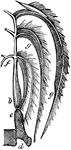
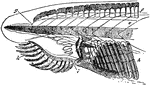
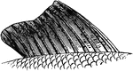
Respiration in Fishes
This figure shows the mode of respiration in fishes. The gills are seen bent over in the form of a feather.…
Fish Skull
"Salmo fario, the entire skull, from the left side. art, articular; branchiost, branchiostegal rays;…
Fish Vertebrae
"Diagram of vertebrae of a bony fish. A, caudal; B, trunk. c, centrum or body of the vertebra; ch.,…
The Muscles of a Fish
In fish the chief masses of the muscular system are disposed on each side of the trunk in a series of…
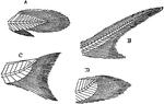
Fishtails
"Diagrams of some principal forms of tails in fishes. A, protocercal fin (as in Polypterus); B, heterocercal…
Gill Arch and Plates
Section through the gill arch and plates of a bony fish. Labels: b, gill plates with capillaries; c,…
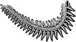
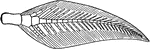
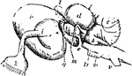
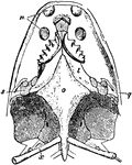
Gills (Branchial Arch of Perch)
This illustration shows the gills (breathing apparatus) of a perch, as well as the specific veins and…
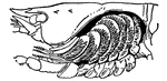
Gills (Crayfish)
This illustration shows the thorax of a crayfish with a portion of the carapace removed to show the…
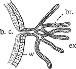
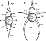
Gills or Branchiae
"Diagram illustrating gills or branchiae. b.c., cavity in which the body fluids circulate; br., branchial…

Parts of Fish Gills
"Gill of Fish. A, first branchial arch of left side of black-bass: 1, gill-rakers; 2, branchial lamellae.…
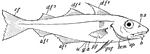
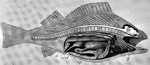
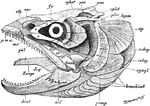
Haddock
"The haddock. n.a., Nasal apertures (double on each side); d.f.1, d.f.2, d.f.3, dorsal unpaired fins;…
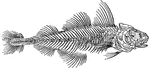
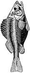
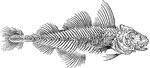
Skeleton of a Haddock
The skeleton of a haddock. In some species such as the haddock, there is a modified form of the coracoid…
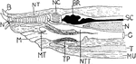
Hagfish Anterior
"Median longitudinal section of anterior region of Myxine. B., Barbule; N., nasal aperture; NT., nasal…
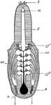
Hagfish Respiratory System
"Respiratory system of hag, from ventral surface. b., Barbules; m., mouth opening on ventral surface;…