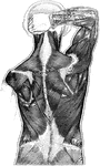
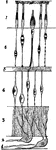
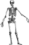
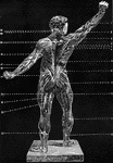
Muscles of the Human Back
Muscles of the back. Labels: 50, latissimus dorsi; 51, trapezius; 52, deltoid. The muscles of the back…
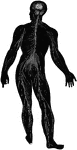
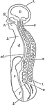
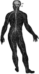
Diagram of the Human Nervous System
Diagram illustrating the general arrangement of the cerebrospinal nervous system.
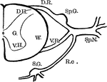
Spinal Nerve Roots
Diagram showing anatomy of the spinal nerve roots and adjacent parts. Labels: G., gray matter of the…
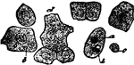
Piece of Human Hair
Piece of human hair, highly magnified. Labels: a, cuticle; b, fibrous substance; c, medulla.
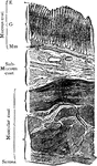
Trunk and Head of Human Body
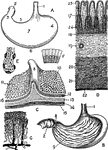
Diagrammatic longitudinal section of the trunk and head. Labels: 1,1, the dorsal cavity; a, the spinal…
Ciliated Epithelium Cells
Ciliated epithelium from the human trachea, highly magnified. Labels: a, large ciliated cell; d, cell,…
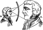
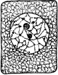
Magnified Face in a Concave Mirror
"When the concave mirror is large, say six inches in diameter, and eight or ten inches focal distance,…
Blood Cells
Red corpuscles (blood cells) of the frog. The red blood cells of birds, reptiles, amphibians and fishes…
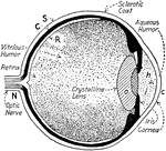
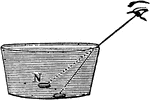
Refraction as seen by the Human Eye
"If the coin were to be observed in an empty pan and then watched as the pan was filled with water,…

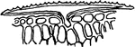
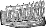
Teeth of an Herbivore
Teeth of an herbivore, showing the rough surface of some of these teeth. Herbivores have no tearing…
Vertebra of a Fish
The vertebra of a fish, which is very different from that of a human. It has but two processes, …
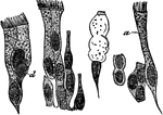
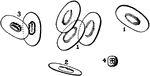
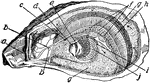
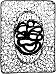
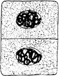
Views of the Stomach
Views of the stomach. Labels: A. stomach (human). B. Same, anterior wall removed. C. Portion of stomach,…
Teeth of Man and Several Animal Species
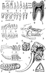
1. Dentition (teeth) of man. 2. Dentition of hyena. 3. Dentition of pig. 4. Dentition of Patagonian…
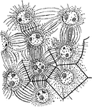
Cell Development
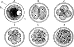
Every human body begin as a single nucleated cell. This cell, known as the ovum, divides or segments…
Retinal Structure
Diagram of the structure of the human retina. Labels: I, pigment layer; II, rod and cone layer; R, rods;…
Teeth
Image of teeth in a human jaw. "1, incisors; 2, canine; 3, bicuspids; 4, molars (the molar at the left…
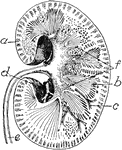
Kidney
Transverse section of the human kidney: "(a) cortex; (b) medulla; (c) small branch of the renal artery;…
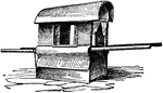
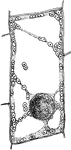
Palanquin
A covered human-powered wagon used in Eastern countries where passengers were inside while two men would…
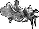
Dinoceras Mirabile
The skull and upper jaw of an early rhinoceros-like mammal from the Cenozoic time.
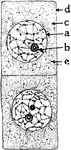
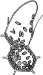
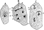
Onion Cells
"A, embryonic cells from onion root tip; d, plasmatic membrane; c, cytoplasm; a, nuclear membrane enclosing…
Onion Cells
"B, older (onion) cells farther back from the root tip. The cytoplasm is becoming vacuolate; f, vacuole."…
T. Zebrina Cell
In onion cells: "C, a cell from the epidermis of the mid-rib of Tradescantia zebrina, in its natural…
T. Majus Cell
"A, cell from the epidermis of the upper side of the calyx of Tropaeolum majus with crystalline chromoplasts."…
Plant Cell Division 1
First stage in plant cell division: Protophase 1; "Resting cell ready to begin division." -Stevens,…
Plant Cell Division 2
Second stage in plant cell division: Protophase 2; "the nuclear reticulum is assuming the form of a…
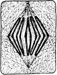
Plant Cell Division 3
Third stage in plant cell division: Protophase 3; "The nuclear thread has divided longitudinally throughout…
Plant Cell Division 4
Fourth stage in plant cell division: Protophase 4; "The nuclear membrane and the nucleolus have disappeared,…
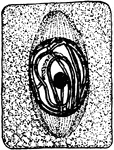
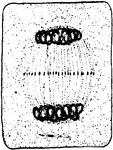
Plant Cell Division 5
Fifth stage in plant cell division: Metaphase; "The metaphase, where the longitudinal halves of the…
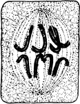
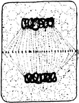
Plant Cell Division 6
Sixth stage in plant cell division: Anaphase; "The anaphase, or movement of the chromosomes toward the…
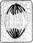
Plant Cell Division 7
Seventh stage in plant cell division: Telophase; "Telophase where the chromosomes have begun to spin…
Plant Cell Division 8
Eighth stage in plant cell division: "The connecting fibers have spread out and come into contact with…
Plant Cell Division 9
Ninth and final stage in plant cell division: "A nuclear membrane has been formed about each daughter…
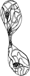
A. Eupatorium Cell
"Formation of endosperm in the embryo-sac of Agrimonia Eupatorium. Cell-walls are being formed between…
E. Communis Cell
"Free cell formation of spores in the ascus of Erysiphe communis. A, ascus with single nucleus; C, cytoplasm;…
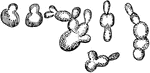
S. Cerevisiae Cell Multiplication
"Various stages of cell multiplication by budding of Saccharomyces cerevisiae." -Stevens, 1916
V. Faba Nucleus Division
"Nucleus dividing by simple constriction. From the lining of the embryo-sac of Vicia faba." -Stevens,…
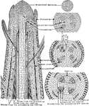
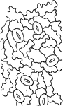
Plant Tissues
"Diagram showing the evolution of tissues from the primordial meristem down to the beginning of cambial…
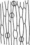
Plant Epidermis
"Two epidermal cells in cross section showing thickened outer wall differentiated into three layers,…