A human skeleton.

Human skeleton

A human skeleton.

Showing the brain cavity of a human.

Human skull

Showing the brain cavity of a human.

"The <em>humerus</em>, a long, hollow bone, rests against a shallow socket on the shoulder blade. It is joined at the elbow to the bones of the forearm." &mdash; Blaisedell, 1904

Humerus

"The humerus, a long, hollow bone, rests against a shallow socket on the shoulder blade. It…
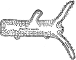
"Longitudinal section of a Hydra; <em>b</em>, bud which will form a young one; <em>ba</em>, base by which it is attached when not creeping." &mdash; Davison, 1906

Section of a hydra

"Longitudinal section of a Hydra; b, bud which will form a young one; ba, base by…
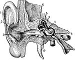
1. Helix 2. Concha 3. Outer passage 4,5,6. emicircular canals 7. Oval indow 8. Cochlea 9. Eustachian tube 10. Ear drum

Inner Ear

1. Helix 2. Concha 3. Outer passage 4,5,6. emicircular canals 7. Oval indow 8. Cochlea 9. Eustachian…
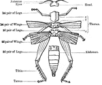
The embryo if an insect, <em>gryllotalpa</em>.

Insect Embryo

The embryo if an insect, gryllotalpa.

"Anatomy of the external skeleton of an insect" &mdash; Goodrich, 1859

External Anatomy of an Insect Skeleton

"Anatomy of the external skeleton of an insect" — Goodrich, 1859

"a, head, antennae, &c; b, pharynx; c, crop; d, gizzard; e, chyle-forming stomach; f, biliary vessels; g, small intestine; h, secreting organs" &mdash; Goodrich, 1859

Digestive Apparatus of an Insect

"a, head, antennae, &c; b, pharynx; c, crop; d, gizzard; e, chyle-forming stomach; f, biliary vessels;…
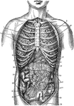
1. Collar bone 2. Left Lung 3. Breast Bone 4. Right Lung 5. Ribs 6. Right lobe of the liver 7. Left lobe of the liver 8. Cartilage 9. Stomach 10. Spleen 11. Descending colon 12. Transverse colon 13. Ascending colon 14. Omentum 15. Coecum 16. Verniform appendix 17.Mesentery 18. Small intestines 19. Sigmoid Flexure 20. Bladder

Internal Anatomy

1. Collar bone 2. Left Lung 3. Breast Bone 4. Right Lung 5. Ribs 6. Right lobe of the liver 7. Left…

"<em>The involuntary muscles</em> consist of ribbon-shaped bands which surround hollow tubes or cavities in the body. They are never attached to bony levers nor are they furnished with tendons, as are the voluntary muscles." — Blaisedell, 1904

Spindle Cell of Involuntary Muscle

"The involuntary muscles consist of ribbon-shaped bands which surround hollow tubes or cavities…

"Showing how the Ends of the Bones are shaped to form the Elbow Joint. The cut ends of a few ligaments are seen." — Blaisedell, 1904

Elbow Joint

"Showing how the Ends of the Bones are shaped to form the Elbow Joint. The cut ends of a few ligaments…

An illustration of a kangaroo pelvis. "M, marsupial bones, borne upon P, pubis; Il, ilium; Is, ischium; O, obturartor foramen; A, acetabulum; S, sacrum;

Kangaroo Pelvis

An illustration of a kangaroo pelvis. "M, marsupial bones, borne upon P, pubis; Il, ilium; Is, ischium;…

"Right wing of the Kestrel, drawn from the specimen, while being held against the light."&mdash;Pettigrew, 1874

Right Wing of Kestrel

"Right wing of the Kestrel, drawn from the specimen, while being held against the light."—Pettigrew,…

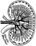
"<em>A</em>, pyramids of the kidney; <em>B</em>, apices, or papillae, of the pyramids; <em>C</em>, pelvis of the kidney; <em>D</em>, upper end of the ureter</em>." &mdash; Blaisedell, 1904

Kidney

"A, pyramids of the kidney; B, apices, or papillae, of the pyramids; C, pelvis…

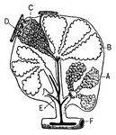
"Diagram of the blood vessels in the kidney." &mdash;Davison, 1910

Kidney

"Diagram of the blood vessels in the kidney." —Davison, 1910

This illustration shows a section on the knee (A, Femur; B, Tibia; C, Patella; D, Synovial sac; E, bursæ). The knee is a hinge-joint, and the bones entering into its formation are the lower end of the femur, the upper end of the tibia, and the posterior surface of the patella (knee-cap).

Section of the Knee

This illustration shows a section on the knee (A, Femur; B, Tibia; C, Patella; D, Synovial sac; E, bursæ).…

"The Lapwing with one wing fully extended, and forming a long lever; the other being in a flexed condition and forming a short lever."&mdash;Pettigrew, 1874

Northern Lapwing

"The Lapwing with one wing fully extended, and forming a long lever; the other being in a flexed condition…
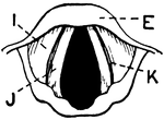
This illustration shows a vertical section of the larynx and its many parts (A. Thyroid Cartilage; B. Cricoid Cartilage; C. hyoid bone; D. Rings of trachea; E. Epiglottis; F. Arytenoid Cartilage; G. Thyro-arytenoid muscle; H. Adam's Apple; I. False Vocal Cords; J. True Vocal Cords; K. Ventricle; L. Rima Glottis).

Vertical Section of Larynx

This illustration shows a vertical section of the larynx and its many parts (A. Thyroid Cartilage; B.…

"Cartilages and Ligaments of the Larynx. (Front view.) <em>A</em>, hyoid bone; <em>B</em>, membrane attached to hyoid bone and the shield-shaped cartilage below (thyroid); edges of this shield-shaped cartilage unite at <em>C</em> (Adam's apple is the V-shaped groove on a line with <em>B</em> and <em>C</em>; <em>D</em>, membrane between the shield-shaped cartilage and the signet-ring cartilage below; <em>E</em> cricoid, or signet-ring, cartilage; <em>F</em>, upper ring of the windpipe." &mdash; Blaisedell, 1904

Front view of the larynx

"Cartilages and Ligaments of the Larynx. (Front view.) A, hyoid bone; B, membrane…
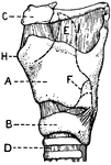
This illustration shows a lateral aspect of the larynx and its multiple parts (A. Thyroid Cartilage; B. Cricoid Cartilage; C. hyoid bone; D. Rings of trachea; E. Epiglottis; F. Arytenoid Cartilage; G. Thyro-arytenoid muscle; H. Adam's Apple; I. False Vocal Cords; J. True Vocal Cords; K. Ventricle; L. Rima Glottis).

Lateral Aspect of Larynx

This illustration shows a lateral aspect of the larynx and its multiple parts (A. Thyroid Cartilage;…

"Cartilages and Ligaments of the Larynx. (Front view.) <em>A</em>, epiglottis; <em>B</em>, thyroid cartilage; <em>C</em>, ladle-shaped cartilage; <em>E</em>, cricoid cartilage; <em>F</em>, upper ring of the windpipe." &mdash; Blaisedell, 1904

Posterior view of the larynx

"Cartilages and Ligaments of the Larynx. (Front view.) A, epiglottis; B, thyroid cartilage;…

This illustration shows the leg of an Ox.
P. Pelvis, FE. Femur, TI. Tibia, FI. Fibula, TA. Tarsus, MT. Metatarsus, PH. Phalanges, OC. Os Calcis.

Leg of Ox

This illustration shows the leg of an Ox. P. Pelvis, FE. Femur, TI. Tibia, FI. Fibula, TA. Tarsus, MT.…

"Muscles of the leg showing how they pass into tendons at the ankle." &mdash;Davison, 1910

Muscles of the leg

"Muscles of the leg showing how they pass into tendons at the ankle." —Davison, 1910

The forelimb of chimpanzee. (c) collar bone; (s) shoulder blade; (h) humerus; (r) radius; (u) ulna; (d) bones of wrist; (m) bones of hand; (p) bones of fingers.

Chimp Limb

The forelimb of chimpanzee. (c) collar bone; (s) shoulder blade; (h) humerus; (r) radius; (u) ulna;…

The hindlimb of chimpanzee. (i) innominate bone; (f) thigh bone; (t) tibia; (s) fibula; (r) bones of ankle; (m) metatarsus; (p) phalanges.

Chimp Limb

The hindlimb of chimpanzee. (i) innominate bone; (f) thigh bone; (t) tibia; (s) fibula; (r) bones of…

Flabellifera are a tribe of isopods. Their bodies end in a tail fan, made by the last pair of appendages and the telson (end piece). While some species of this tribe are parasitic, this particular species, Limnoria Lignorum, is non-parasitic, and has a destructive action on submerged wood. It is commonly referred to as the 'gribble'.

Limnoria Lignorum (Flabellifera)

Flabellifera are a tribe of isopods. Their bodies end in a tail fan, made by the last pair of appendages…

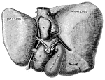
"The Liver seen from Below and Behind. <em>A</em> and </em>B</em>, smaller lobes of the liver; <em>C</em>, portal vein; <em>D</em> hepatic artery; <em>E</em>, inferior vena cava; <em>F</em>, trunk of the left hepatic vein; <em>G</em>, hepatic vein." &mdash; Blaisedell, 1904

Liver

"The Liver seen from Below and Behind. A and B, smaller lobes of the liver; C,…

External features of the lobster.

Lobster

External features of the lobster.

"Section of a pulmonary lobule, showing its division into pulmonary vesicles." &mdash; Tracy, 1888

Pulmonary lobule

"Section of a pulmonary lobule, showing its division into pulmonary vesicles." — Tracy, 1888

"Little openings (Haversian canals) are seen, and around them are arranged rings of bone with little dark spaces (<em>lacunae</em>), from which branch out fine dark lines (<em>analiculi</em>)." &mdash; Blaisedell, 1904

Cross-section from a shaft of a long bone

"Little openings (Haversian canals) are seen, and around them are arranged rings of bone with little…

"<em>A</em>, an air sac; <em>B</em>, an air sac cut open; <em>C</em>, capillary network over an air sac; <em>D</em>, branch of pulmonary artery; <em>E</em>, branch of pulmonary vein; <em>F</em>, bronchial tube." &mdash; Blaisedell, 1904

Lobule of the lung

"A, an air sac; B, an air sac cut open; C, capillary network over an air…

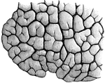
"Lobules of various sizes are well shown." &mdash; Blaisedell, 1904

Portion of a child's lung

"Lobules of various sizes are well shown." — Blaisedell, 1904

In the lungfish, the development of the air bladder as a lung is much more complete than others in the same group.

Lungfish

In the lungfish, the development of the air bladder as a lung is much more complete than others in the…
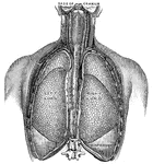
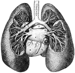
"<em>L.A.</em>, left ventricle; <em>B</em>, right ventricle; <em>F</em>, pulmonary artery; <em>G</em>, aorta; <em>H</em>, arch of the aorta; <em>K</em>, innominate artery; <em>L</em>, right common carotid artery; <em>M</em>, right subclavian artery; <em>N</em>, thyroid cartilage forming upper portions of the larynx; <em>O</em>, trachea." &mdash; Blaisedell, 1904

Lungs

"L.A., left ventricle; B, right ventricle; F, pulmonary artery; G,…

"<em>A</em>, division of left bronchus to upper lobe; <em>B</em>, left branch of the pulmonary artery; <em>C</em>, left bronchus; <em>D</em>, left superior pulmonary vein; <em>E</em>, left inferior pulmonary vein; <em>F</em>, left auricle; <em>K</em>, inferior vena cava; <em>L</em>; division of the right bronchus to lower lobe; <em>M</em>, right inferior pulmonary vein; <em>N</em>, right superior pulmonary vein, <em>O</em>, right branch of the pulmonary artery; <em>P</em>, division of right bronchus to upper lobe; <em>R</em>, left ventricle; <em>S</em>, right ventricle." &mdash; Blaisedell, 1904

Lungs

"A, division of left bronchus to upper lobe; B, left branch of the pulmonary artery;…

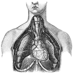
"Relative Postion of the Lungs, the Heart, and Some of the Great Vessels belonging to the latter. <em>A</em>, left common carotid artery; <em>B</em>, external carotid artery; <em>C</em>, internal carotid artery; <em>D</em>, left jugular vein; <em>E</em>, sterno-cleido muscle; <em>F</em>, right innominate artery with branches to head and arm; <em>G</em>, left subclavian artery; <em>H</em>, great azygos vein; <em>K</em>, thoracic duct." &mdash; Blaisedell, 1904

Lungs

"Relative Postion of the Lungs, the Heart, and Some of the Great Vessels belonging to the latter. A,…
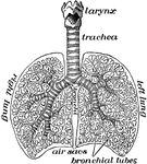
"The lungs fill up most of the cavity of the chest. One lies on either side of the heart which is in the middle of the chest. The lungs in animals are called lights because they are spongy sacs and so light as to float when thrown into water." &mdash;Davison, 1910

The lungs

"The lungs fill up most of the cavity of the chest. One lies on either side of the heart which is in…

"The lymph vessels of the body. rc, the thoracic duct; lac, the lacteals taking the lymph and fatty part of food from the intestines." —Davison, 1910

Lymph Vessels

"The lymph vessels of the body. rc, the thoracic duct; lac, the lacteals taking the lymph and fatty…

"Consisting of a very fine network of fibrils, around which are cells of various sizes." — Blaisedell, 1904

Connective Tissue from a Lymphatic Gland

"Consisting of a very fine network of fibrils, around which are cells of various sizes." — Blaisedell,…

The perch is typical of a large group of fishes, all of which have spiny rays. The perch is widely distributed in fresh-water lakes and streams.

Mackerel

The perch is typical of a large group of fishes, all of which have spiny rays. The perch is widely distributed…
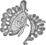
S, cut end of duodenum; R, cut end of rectum; C, caecum; C2, accessory caecum; C.L., colic loop of hind-gut.

Intestinal Tract of Macropus Bennetti

S, cut end of duodenum; R, cut end of rectum; C, caecum; C2, accessory caecum; C.L., colic loop of hind-gut.

Megatherium ("Great Beast") was a genus of elephant-sized ground sloths that lived from two million to 8,000 years ago. A related genus was Nothrotheriops, which were primarily bear-sized ground sloths. The rhinoceros-sized Promegatherium is suggested to be the ancestor of Megatherium.

Megatherium Skeleton

Megatherium ("Great Beast") was a genus of elephant-sized ground sloths that lived from two million…

"A Compound Microscope. The appearance of the various structures and tissues of the human body as revealed by the microscope possesses a curious fascination for every observer, especially for young students. No one ever forgets the first look at a drop of blood, or the circulation of blood in the web of a frog's foot as revealed by the microscope." &mdash; Blaisedell, 1904

Compound microscope

"A Compound Microscope. The appearance of the various structures and tissues of the human body as revealed…
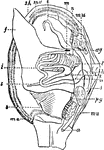
"Anatomy of an Acephalous Mollusc (Mactra): s, stomach; ii, intestine; ag, anterior ganglions; pg, posterior ganglions; mn, muscles; a, anus; h, heart; l, liver; f, foot; sh, shell; ma, mantle; b, branchiae; t, tentacula; r, oral, or respiratory syphon; t, anal syphon." &mdash; Chambers, 1881

Mollusc Anatomy

"Anatomy of an Acephalous Mollusc (Mactra): s, stomach; ii, intestine; ag, anterior ganglions; pg, posterior…

An illustration of the mouth organs of the stag beetle.

Mouth Organs of a Beetle

An illustration of the mouth organs of the stag beetle.

"A muscle cell from the stomach." &mdash; Ritchie, 1918

Muscle cell

"A muscle cell from the stomach." — Ritchie, 1918

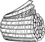
"Fine threads of fibers forming a muscle." &mdash;Davison, 1910

Muscle fibers

"Fine threads of fibers forming a muscle." —Davison, 1910

"Principal Muscles on the Right, Certain Organs of the Chest and Abdomen, and the Larger Blood Vessels on the Left." — Blaisedell, 1904

Muscles and Blood Vessels

"Principal Muscles on the Right, Certain Organs of the Chest and Abdomen, and the Larger Blood Vessels…

"The extensor muscles on the back of the forearm. Note the tendons at the wrist." &mdash;Davison, 1910

Muscles of the forearm

"The extensor muscles on the back of the forearm. Note the tendons at the wrist." —Davison, 1910

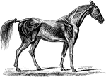
The superficial muscles in a horse.

Horse Muscles

The superficial muscles in a horse.

"<em>A</em>, section of cutaneous fold turned back to show root of nail; <em>B</em>, cutaneous fold covering root of nail; <em>C</em>, semilunar whitish portion; <em>D</em>, body of nail." &mdash; Blaisedell, 1904

Nail in position

"A, section of cutaneous fold turned back to show root of nail; B, cutaneous fold…

"Concave or Adherent Surface of the Nail. <em>A</em>, border of the root; <em>B</em>, whitish portion of semi-lunar shape; <em>C</em>, body of nail. The continuous line around border represents the free edge." &mdash; Blaisedell, 1904

Surface of a nail

"Concave or Adherent Surface of the Nail. A, border of the root; B, whitish portion…

"Diagram of a Sectional View of Nasal and Throat Passageways. <em>C</em>, nasal cavities; <em>T</em>, tongue; <em>L</em>, lower jaw; <em>M</em>, Mouth; <em>U</em>, uvula; <em>E</em>, epiglottis; <em>G</em>, larynx; <em>O</em>, gullet, or esophagus." &mdash; Blaisedell, 1904

Nasal and throat passageways

"Diagram of a Sectional View of Nasal and Throat Passageways. C, nasal cavities; T,…
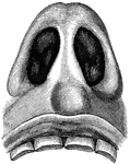
"Nasal Cavities, seen from Below. The sense of smell is located in the membrane which lines the cavities of the nose. This delicate membrane, over which the fibers of the olfactory nerves, or the nerves of smell, are distributed, is kept continually mois by the mucus which it secretes." &mdash; Blaisedell, 1904

Nasal cavities

"Nasal Cavities, seen from Below. The sense of smell is located in the membrane which lines the cavities…

An illustration of a female nautilus without the shell. "m, The dorsal "hood" formed by the enlargement of the outer or annular lobe of the forefoot, and corresponding to the sheaths of two tentacles; n. Tentacular sheaths of lateral portion of the annular lobe; u, The left eye; b, The nuchal plate, continuous at its right and left posterior angles with the root of the mid-foot, and corresponding to the nuchal cartilage of Sepia; c, Visceral hump; d, The free margin of the mantle-skirt,...; g.a, Points to the lateral continuation of the nuchal plate b to join the root of the mid foot of siphon." (Britannica, 1910).

Female Nautilus without Shell

An illustration of a female nautilus without the shell. "m, The dorsal "hood" formed by the enlargement…

An illustration of a female nautilus without the shell. "c, points to the concave margin of the mantle-skirt leading into the sub-pallial chamber; g, the mid-foot or siphon; k, the superficial origin of its retractor muscles closely applied to the shell and serving to hold the animal in tis place; l, the sipjuncular pedicle of the visceral hump broken off short; v,v, the superior and inferior ophthalmic tentacles." (Britannica, 1910)

Female Nautilus without Shell

An illustration of a female nautilus without the shell. "c, points to the concave margin of the mantle-skirt…
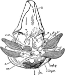
An illustration of the postero-ventral view female nautilus without the shell. "a, Muscular band passing from the mid-foot to the integument; b, The valve on the surface of the funnel, partially concealed by the inrolled lateral margin of the latter; c, The mantle-skirt retroverted; an, The median anus; x, Post-anal papilla of unknown significance; g.n., Nidamental gland ; r.ov, Aperture oft he right oviduct; l.ov, Aperture of the rudimentary left oviduct; neph.a, Aperture of the left anterior renal sac; neph.p, Aperture of the left posterior renal sac; viscper, Left aperture of the viscero-pericardial sac; olf, The left osphradium placed near the base of the anterior gill-plume." (Britnnica, 1910)

Female Nautilus without Shell

An illustration of the postero-ventral view female nautilus without the shell. "a, Muscular band passing…
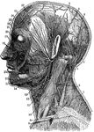
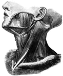
1. Temporal Artery 2. Artery behind the ear 3. Occipital Artery 4. Greater occipital nerve 5. Smaller ocipital nerve 6. Nerve of the neck 7. Trapezius muscle 8. Clavicular nerve 9. Clavicle 10. Sterno-0cleido-mastoid muscle 11.Outer artery of the head 12. Inner artery of the head 13. Salivary gland 14. Nerves of the lower jaw 15. Outer maxillary artery 16. Nerve of the chin 17. Circular muscle of th emouth 18. Greater yoke muscle 19. Nerves below the eye 20. Masseter or chewing muscle 21. Ear passage 22. Arteries of the forehead 23. Nerves of the forehead 24. Eye closing muscle 25.Facial Artery 26. Facial nerve

Neck

1. Temporal Artery 2. Artery behind the ear 3. Occipital Artery 4. Greater occipital nerve 5. Smaller…

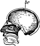
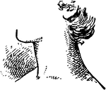
The side view of a human neck.

Neck

The side view of a human neck.

"Principal Muscles on the Left Side of the Neck." &mdash; Blaisedell, 1904

Muscles of the neck

"Principal Muscles on the Left Side of the Neck." — Blaisedell, 1904