"Disarticulation of bird's skull. Membrane bones shaded. B.Oc., basioccipital; E.Oc., exoccipital; S.Oc., supraoccipital; Pa., parietal; Fr., frontal; Na., nasal; pm., premaxilla; M., maxilla; Ju., jugal; Qj., quadrato-jugal; Qu., quadrate; pe., periotic; Sq., squamosal; AS., alisphenoid; B.S., basisphenoid; O.S., orbito-sphenoid; Pr.Sph., presphenoid; vo., vomer; iOS., interorbitalk septum; E., ethmoid; Se., nasal septum; De., dentary; Sp., splenial; An., angular; Ar., articular; MK., Meckel's cartilage." -Thomson, 1916

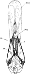
Bird Skull Disarticulation

"Disarticulation of bird's skull. Membrane bones shaded. B.Oc., basioccipital; E.Oc., exoccipital; S.Oc.,…
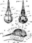
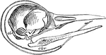
"Fig 66 - Head of a chick, second stage, after five days of incubation, section in profile; x6 diameters. cvl, cv2, cv3, first, second, and third cerebral vesicles; 1, place of the first nerve, the olfactory; 2, place of second nerve, the optic; ic, internal carotid artery, running into skull at what was originally the pituitary space, now an opening bounded in front by the anterior, acl, behind the posterior, pcl, clinoid walls; nc, notochord; oc, occipital condyle, thence to pcl being the original parachordal cartilage, here seen in profile; eo, exoccipital; eth, ethmoid, with ps, its presphenoid region posteriorly, and pn, pre-nasal part; this whole plate afterward developing into parts of the nose and the partition between the eyes; pa, palatine; pg, pterygoid region; pa and pg reference lines are in the chick's mouth; mk meckelian cartilage (lower jaw); ch and bh, ceratohyal and basihyal parts of the hyoid or tongue bone." Elliot Coues, 1884

Chick Head

"Fig 66 - Head of a chick, second stage, after five days of incubation, section in profile; x6 diameters.…

"Phalacrocorax bicristatus. Red-Faced Cormorant. Skull showing sto, occipital style or nuchal bone; The style is somewhat tilted upward from its natural position." Elliot Coues, 1884

Cormorant Skull

"Phalacrocorax bicristatus. Red-Faced Cormorant. Skull showing sto, occipital style or nuchal bone;…
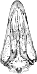
"Schizognathous skull of common fowl, nat. size, from nature, by Dr. R.W. Shufeldt, U.S.A. Letters as before; Pa, palatine. Schizognathism is a kind of " cleft palate" shown by the columbine and gallinaceous birds, by the wader at large, and many of the swimmers. In this general case, the vomer, whether large or small, tapers to a point in front, while behind it embraces the basisphenoidal rostrum, between the palatines; these bones and the pterygoids are directly articulated with one another and with the basisphenoidal rostrum, not being borne upon the divergent posterior ends of the vomer; the maxillo-palatines, usually elongated and lamelar, pass inwards over (under, when the skull is viewed upside-down, as it usually is) the anterior part of the palatines, with which they unite and then bend backwards, along the inner edge of the palatines, leaving a broader or narrower fissure between themselves and the vomer, on each side, and do not unite with one another or with the vomer." Elliot Coues, 1884

Common Fowl Skull

"Schizognathous skull of common fowl, nat. size, from nature, by Dr. R.W. Shufeldt, U.S.A. Letters as…

"Dromaeognathous skull of ostrich, nat. size specimen no. 16,629, U.S. Nat Museum, by Dr. R. W. Shufeldt, U.S.A. RR, rostrum, beyond which the ossified nasal septum continues in the axis of the skull to the letters "Pmx." V, the short vomer, borne upon R, uniting laterally with Mxp, the broad maxillo-palatines; Pl, palatines, remote from rostrum, underrunning beyond Mxp, but not to Pmx. Pt, expanded scroll-like pterygoids, atriculating behind with Btp, the strong basipterygoid processes on the body (not rostum) of the sphenoid; they underlap R, but do not articulate there. Pmx, premaxillaries; Mx, maxillaries, whose ends run forward to opposite the letters "Pmx"; jugal; qj, quadrato-jugal; Qu, quadrate. (N.B. This is the most exceptional case of dromaeognathism. Each of the Ratite families - Struthionidae, Rheidae, Casuariidae, Dinornithidae, and Apterygidae, - as well as the Carinate family Tinamidae, offers a special case of such formation, as explained in the text.)." Elliot Coues, 1884

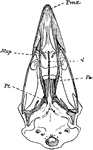
The Skull Structure of an Ostrich

"Dromaeognathous skull of ostrich, nat. size specimen no. 16,629, U.S. Nat Museum, by Dr. R. W. Shufeldt,…
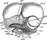
"Columba livia. Skull of young specimen. A, dorsal; B, ventral; C, left side. al. s, alisphenoid; au, angular; ar, articular; b. o, basi-occipital; d, dentary; e. o, ex-occipital; eu, aperture of Eustachian tube; f. m, foramen magnum; fr, frontal; i.o. s, inter-orbital septum; ju, jugal; lc, lachrymal; lb. s, lambdoidal suture; m. eth, mesethmoid; mx, maxilla; mx. p, maxillo-palatine process; na, na', na'', nasal; o. c, occipital condyle; or. fr, orbital plate of frontal; pa, parietal; pa. s, parasphenoid (rostrum); pl, palatine; p. mx, premaxilla; pt, pterygoid; qu, quadrate; s. an, supra-angular; s. o, supra-occipital; sq, squamosal; ty, tympanic cavity; IL-XII, foramina for cerebral nerves." -Parker, 1900

Rock Pigeon Skull

"Columba livia. Skull of young specimen. A, dorsal; B, ventral; C, left side. al. s, alisphenoid; au,…

"Aegithognathous skull of raven, Corvus corax, nat. size, from nature, by Dr. R. W. Shufeldt, U.S.A. Letters as before. N.B. The reference line, V, goes to the ossified nasal septum borne upon the end of the vomer, which latter bone begins at the thickest part of the central projection. Mxp underlies V and overlies Pl, but touches neither." Elliot Coues, 1884

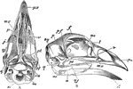
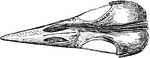
The Skull of a Raven

"Aegithognathous skull of raven, Corvus corax, nat. size, from nature, by Dr. R. W. Shufeldt, U.S.A.…
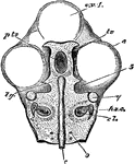
The skull of a bird. Labels: a, inferior aspect, the mandible being removed; b, lateral aspect; px, premaxilla; mx, superior maxilla; p, palatine bone; v, vomer; j, jugal bone; qj, quadrato-jugal bone; lachrymal; me, median ethmoid; f, frontal; pf, post-frontal process; p, parietal; pt, pterygoid; q, os quadratum; sq, sqamosal; eu, process before the sustachian aperture; bo, basi-occipital, and so, supra-occipital bone; ar, articular; a, surangular, and d, dentary portion of the mandible.

Skull of a Bird

The skull of a bird. Labels: a, inferior aspect, the mandible being removed; b, lateral aspect; px,…

"Fig 64 - Skull of chick, fifth day of incubation, x 9 diameters. Seen from above, the membranous roof of the skull and the brain removed. cv1, anterior cerebral vesicle; e, eye; c, notochord, running through the middle of the basilar plate or parachordal cartilage, in which are already visible the rudimentary ear-parts, cl, the cochlea, hsc, the horizontal semicircular canal; pts, the pituitary space, bounded by tr, the trabeculae, which come together before it to form the fronto-nasal plate, fn, in fig. 65; lg, lingula or bridge connecting trabeculae with parachordal cartilage; 5 notch afterward becoming foramen ovale for passage of parts of the fifth (trifacial) nerve; 9, foramen for hypoglossal nerve; q, separate cartilage forming the future quadrate bone." Elliot Coues, 1884

Skull of a Chick

"Fig 64 - Skull of chick, fifth day of incubation, x 9 diameters. Seen from above, the membranous roof…

"Skull of a chick, but seen from below. cv1, anterior cerebral vesicle; e, eye; m, mouth; pts, pituitary space; fn, fronto-nasal plate; tr, ends of the trabeculae, free again after their union and bent strongly from the original axis of the trabeculae; n, external nostril; mxp, subocular bar of cartilage, or pterygo-palatine rod, to form pa, palatine, and pg, pterygoid bone, and other parts of the upper jaw, as the maxillary, jugal and quadrato-jugal; q, quadrate cartilage, same as seen in fig 64; mk, meckelian cartilage, to form lower jaw; these parts are in the first post-oral visceral arch; ch, cerato-hyal, and bh, basihyal, of second postoral arch; cbr, cerato-branchial, ebr-branchial, bbr, basi-branchial, of third post-oral arch; the parts of the second and third arch all going into the hyoid bone. 1, 2, 3, 1st, 2d, 3d, visceral clefts, whereof the 1st is to be modified into the ear-passages, and the others are to be obliterated." Elliot Coues, 1884

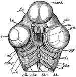
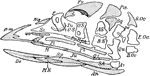
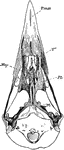
Skull of a Chick Below

"Skull of a chick, but seen from below. cv1, anterior cerebral vesicle; e, eye; m, mouth; pts, pituitary…

"Side view of a woodpecker's skull, showing the long slender basihyal (bh), bearing slight elements at its fore end, no uroyhal, and extraordinarily long thyrohyals (cbr, ebr) curving up over back of skull and curling together around orbit of the right eye." Elliot Coues, 1884

The Skull of a Woodpecker

"Side view of a woodpecker's skull, showing the long slender basihyal (bh), bearing slight elements…
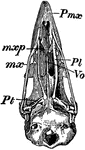
"Schizognathous skull of common fowl. pmx, premaxilla; mxp, maxillopalatine; mx, maxilla; pl, palatine; pt, pterygoid; vo, vomer." -Whitney, 1911

Bird Skull

"Schizognathous skull of common fowl. pmx, premaxilla; mxp, maxillopalatine; mx, maxilla; pl, palatine;…
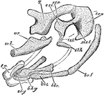
"The post-oral arches of the house martin, at middle of period of incubation, lateral view, X14 diameters. mk, stumpof meckelian or mandibular rod, its articular part, ar already shapen; q, quadrate bone, or suspensorium of lower jaw, with a free anterior orbital process and long posterior otic process articulating with the ear-capsule, of which teo, tympanic wing of occipital, is a part; mst, est, sst, ist, sth, parts of suspensorium of the third post-oral arch, not completed to chy; mst, medio-stapedial, tp come away from teo, bringing a piece with it, the true stapes or columella auris; the oval base of the stapes fitting into the future fenestra ovalis, or oval window looking into the cochlea; sst, supra-stapedial; est, extra-stapedial; ist, infra-stapedial, which will unite with sth, the stylo-hyal; chy and bhym cerato-hyal and basi-hyal, distal parts of the same arch; bbr, br 1, br2, basi-branchial, epi-branchial and cerato-branchial pieces of the third arch, composing the rest of the hyoid bone; tg, tongue." Elliot Coues, 1884

House Martin Skull

"The post-oral arches of the house martin, at middle of period of incubation, lateral view, X14 diameters.…

"Desmognathous skull of mallard duck, Anas boscas, nat. size, from nature, by Dr. R.W. Shufeldt, U.S.A. Letters before. In the "bound-palate" type, the vomer in either abortive, or so small that it disappears; when existing it is usually slender and tapers to a point in front; the maxillo-palatines are united across the median line, either directly or by means of ossification in the nasal septum; the posterior ends of the palatines and the anterior ends of the pterygoids articulate directly with the rostrum (as in schizognathism). This type is simply and perfectly exhibited by a duck in which the maxillo-palatine is a broad flat plate united with its fellow in mid-line; the oval sessile basipterygoid facets are far forward, opposite the very ends of the pterygoids." Elliot Coues, 1884

Mallard Duck Skull

"Desmognathous skull of mallard duck, Anas boscas, nat. size, from nature, by Dr. R.W. Shufeldt, U.S.A.…

"Dromaeognathous skull of a tinamou (Tinamus robustus); copies by Shufeldt from Huxley. Letters as before; Mxp, maxillo-palatine. The tinamous, Dromaeognathae "have a completely struthious palate"; vomer very broad, uniting in front with broad maxillo-palatine plates as in Dromaeus; behind articulating with posterior ends of palatines and anterior ends of pterygoids, both of which are thus prevented, as in all Ratitae, from any extensive connection with the rostrum; basipterygoid processes springing from body of sphenoid, not from its rostrum, articulating with pterygoids very near the posterior or outer ends of the latter; head of quadrate with a single articular facet, as in Ratitae." Elliot Coues, 1884

The Skull of a Tinamou

"Dromaeognathous skull of a tinamou (Tinamus robustus); copies by Shufeldt from Huxley. Letters as before;…

"Top view of skull of Cloaptes, (flickers) showing thyrohyals running along the skull and into right nostril to end of the bill." Elliot Coues, 1884

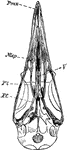
Top View of a Woodpecker Skull

"Top view of skull of Cloaptes, (flickers) showing thyrohyals running along the skull and into right…