Clipart tagged: ‘bird tongue’
Chick Head
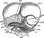
"Fig 66 - Head of a chick, second stage, after five days of incubation, section in profile; x6 diameters.…
Bustard Gular Pouch
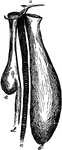
"Gular pouch of bustard; a, tongue; b, the pouch, opening under a, hanging in front of c, the trachea,…