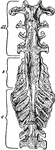
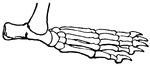
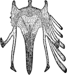
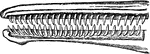
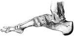
Bones of the Ankle and Foot
Bones of the Ankle and Foot. Labels: m, metatarsal bones; p, phalanges; ca, os calcis, or heel bone.
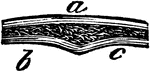
Arm Bones
"The bones of the arm. a, humerus; b, ulna; c, radius; d, the carpus; e, the fifth metacarpal; f, the…
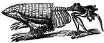
Armadillo - Endoskeleton and Exoskeleton or Dermoskeleton
An illustration of a pichiciago, a small burrowing armadillo. The front half of the animal is covered…
Artiodactyles
"Bones of fore foot of existing Artiodactyle. Pig (Sus scrofa)." —The Encyclopedia Britannica,…
Artiodactyles
"Bones of fore foot of existing Artiodactyle. Red Deer (Cervus elaphus)." —The Encyclopedia Britannica,…
Leg of Bear
This illustration shows the plantigrade leg of a bear. Plantigrade means that the animal walks flat…
Wing of Bird
"Shows how the bones of the arm (a), forearm (b), and hand (c), are twisted, and form a conical screw."—Pettigrew,…
Bone Exposed to Acid and Twisted
This figure shows a thigh bone that has been softened by exposing it to acid, then twisted in a knot…
Formation of Compact Bone in a Human
Transverse section of femur of a human embryo about eleven weeks old. Labels: a, rudimentary Haversian…
Formation of Compact Bone in a Kitten
Transverse section through the tibia of a fetal kitten. Labels: P, Periosteum. O, Osteogenetic layer…
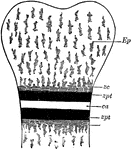
Bone Ossification
Schematic diagram, showing epiphysis and diaphysis and line of ossification. Labels: Ep, epiphysis of…
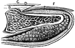
Bone Structure
If we divide any of the long bones longitudinally, we find two kinds of structure, the hard or compact,…
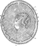
Bone Tissue of Humerus
Transverse section of compact tissue of humerus, magnified about 150 diameters. Three of the Haversian…
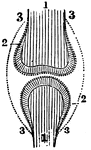
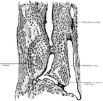
Position of the Bone, Cartilage, and Synovial Membranes
A diagram of the relative position of the bone, cartilage, and synovial membrane. Labels: 1,The extremities…
Developing Bone
Cross section of developing bone of human fetus of four months. Labels: a, periostem; b, boundary between…
A Magnified View of a Bone
A thin slice of bone, highly magnified, showing the lacunae, the tiny tubs (canaliculi) radiating from…
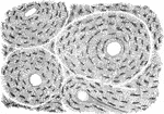
Microscopic Structure of Bone
The bone contains a multitude of small irregular spaces, approximately fusiform in shape, called lacunae,…
Microscopic Structure of Bone
The bone contains a multitude of small irregular spaces, approximately fusiform in shape, called lacunae,…
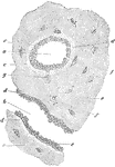
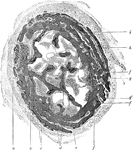
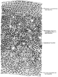
Section of Bone
A small piece of bone, ground very thin and highly magnified. "If a bit of bone is still more magnified…
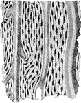
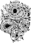
Transverse Section of Compact Bone
Transverse section of compact bone (metatarsal); the section has been ground and dried, hence the lacunae…
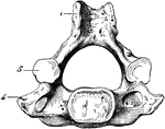
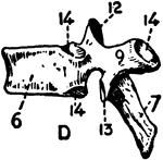
Human Cervical Vertebra Bone
A cervical vertebra of the spine, inferior surface. Labels: 1, spinous process, slightly bifid; 4, transverse…
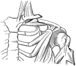
Broken Clavicle
"When a bone is broken, blood trickles out between the incjured parts, and afterwards gives place to…
Compact Bone
"Compact bone consists of a series of concentric layers of bone disposed around a canal called the Haversian…
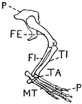
Leg of Crocodile
This illustration shows the leg of a crocodile. P. Pelvis, FE. Femur, TI. Tibia, FI. Fibula, TA. Tarsus,…
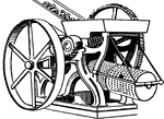
Cross Mill and Sieves (Glue)
This illustration shows a cross mill and the sieves used to crush and filter bones in glue manufacturing.
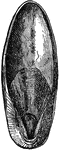
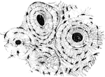
Cuttlebone
Cuttlefish "bone" or internal shell. The fine point at the base structure represents the guard of the…
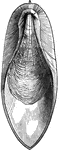
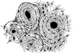
Bone of the cuttle-fish
"An internal support of a calcareous nature, and formed in laminae; this is the well-known cuttlefish…
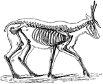
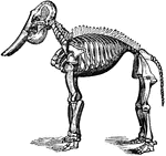
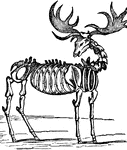
Skeleton of the Deer
"The bones in the extremities of this the fleetest of quadrupeds are inclined very obliquely towards…
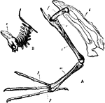
Diver Bones
"A. Pelvis and bones of the leg of the Leon or Diver; i, Innominate bone; f, Thighbone (femur); r, Tibia;…
Leg of Dog
This illustration shows the leg of a dog. This leg is digitigrade. Animals with digitigrade legs walk…
Dolphin Teeth
"Upper and lower teeth of one side of the mouth of a dolphin, as an example of homodont type of dentition.…
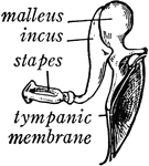
Bones of the Ear
"Across the middle ear a chain of three small bones stretches from the tympanic membrane to the inner…
Bones of the Ear
"1, malleus, or hammer; 2, incus, or anvil; 3, stapes, or stirrup." — Blaisedell, 1904
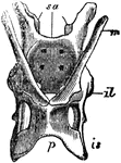
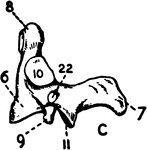
Echidna Pelvis
"The pelvis of the Echidna; sa, sacrum; il, illum; is, ischium; p, pubis; m, marsupial bone." —…
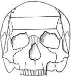
Ethmoid Bone of the Human Skull
Ethmoid bone, posterior surface. The ethmoid bone is an exceedingly light, spongy bone, placed between…
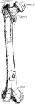
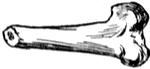
Human Femur Bone
The Femur (upper leg bone) is the longest, largest, and strongest bone in the skeleton. Labels: b, rounded…
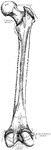
Anterior View of Human Right Femur
"Anterior View of Human Right Femur. ec, external condyle; etu, external tuberosity; ic, internal condyle;…
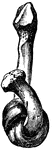
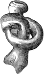
Softened fibula
"The Fibula tied into a Knot after the Mineral Matter has been dissolved by Acid." — Blaisedell,…
Longitudinal seciont of a fingernail
"A, last bone of finger; B, true skin on the dorsal surface of finger; C,…
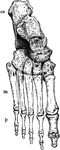
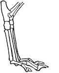
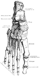
Ligaments of the Foot and Ankle
"The bones are fastened together, kept in place, and their movements limited, by tough and strong bands,…
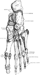
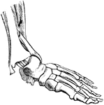
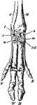
Bones of the Foot
"The foot is built in the form of a half-dome or half-arch. This is to afford a broad, strong support…