Clipart tagged: ‘"cervical vertebrae"’
Different View of the Spinal Cord
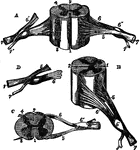
Different views of a portion of the spinal cord from the cervical region, with roots of the nerves slightly…
Different views of a portion of the spinal cord from the cervical region, with roots of the nerves slightly…