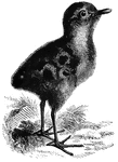
Clipart tagged: ‘chick’
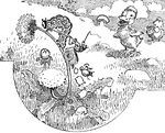
Chick Head
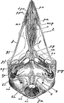
"Fig 66 - Head of a chick, second stage, after five days of incubation, section in profile; x6 diameters.…
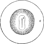
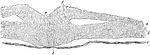
Vascular Area in the Chick
Diagram showing vascular area in the chick. A, area pellucida; b, area vasculosa; c, area vitellina.
Embryo Chick
Embryo chick (36 hours), viewed from beneath as a transparent object (magnified). Labels:pl, outline…
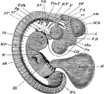
Embryo Chick at Fourth Day
Embryo chick at fourth day, viewed as a transparent object, lying on its left side. CH, cerebral hemispheres;…
Cells of an Embryo Chick
A transverse section through an embryo chick (26 hours). Labels: a, epiblast; b, mesoblast; c, hypoblast;…
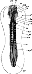
Transverse Section of an Embryo Chick
Transverse section of an embryo chick (third day). Labels: mr, rudimentary spinal cord; the primitive…
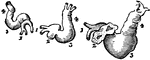
Development of the Heart of a Chick
Heart of a chick at the 45th, 65th, and 85th hours of incubation. I, the venous trunks; 2, the auricle;…
Intestine of a Chick
Rudiments of the liver on the intestines of a chick at the fifth day of incubation. Labels: 1, heart;…
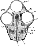
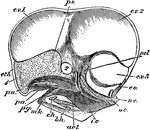
Skull of a Chick
"Fig 64 - Skull of chick, fifth day of incubation, x 9 diameters. Seen from above, the membranous roof…
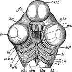
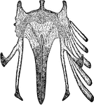
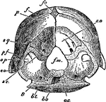
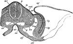
Skull of a Chick Below
"Skull of a chick, but seen from below. cv1, anterior cerebral vesicle; e, eye; m, mouth; pts, pituitary…