Clipart tagged: ‘"ciliary muscle"’
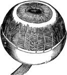
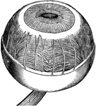
Left Eyeball in Horizontal Section
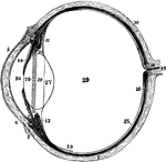
The left eyeball in horizontal section from before back. Labels: 1, sclerotic; 2, junction of sclerotic…
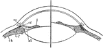
Focusing of the Eye
Diagram to illustrate the mechanism of accommodation (focusing); on the right half of the figure for…