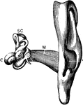
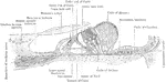
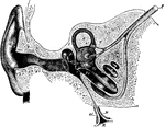
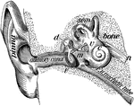
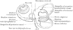
The Auditory Apparatus
The auditory apparatus with the surrounding bone removed. Labels: M, external auditory canal; SC, semicircular…
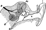
Diagram of the Auditory Apparatus
A diagram of a section of the auditory apparatus. Labels: E, external canal; M, in the middle ear; V,…
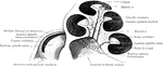
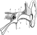
View of the Auditory Nerve
A view of the auditory nerve. Labels: 1, The spinal cord. 2, The medulla oblongata. 3, The lower part…
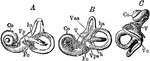
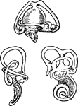
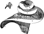
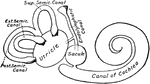
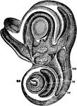
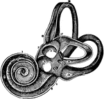
The Bony Labyrinth of the Ear
Casts of the bony labyrinth of the ear. Labels: A, left labyrinth see from the outer side; B, right…

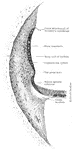
External auditory canal
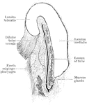
"A Cast of the External Auditory Canal. The auditory canal is a passage in the solid potion of the temporal…
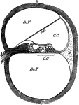
Cochlea
Diagram of a section of a coil of the cochlea of the ear. Labels: C.C, canal of the cochlea; mR, its…
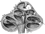
The Cochlea of the Ear
A vertical section of the cochlea, highly magnified, to show the arrangement and connection of its parts.
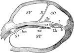
Cochlea, Section of
Section of one coil of the cochlea, magnified. Labels: SV, scala vestibuli; R, membrane of Reissner;…
Ear
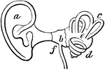
Interior of the ear. There is external to the head a wide-mouthed tube, or ear-trumpet (a), for catching…
Ear
The human ear. Labels: A, vestibule or antechamber; B, auditory canal; C, the hammer, anvil, and the…
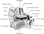
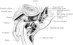
Ear and Auditory Canal
Semi-diagrammatic section through the right ear. Labels: M, concha; G, the external auditory canal;…
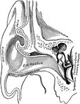
Ear Showing External Auditory Meatus
Vertical section through the external auditory meatus and tympanum, passing in front of the fenestra…
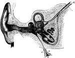
Auditory Apparatus of the Ear
Horizontal section through the right external acoustic canal viewed from above.
Auditory Canal of the Ear
Vertical section through the right external acoustic canal, viewed from in front.
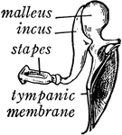
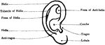
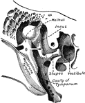
Bones of the Ear
"Across the middle ear a chain of three small bones stretches from the tympanic membrane to the inner…
Bones of the Ear
"1, malleus, or hammer; 2, incus, or anvil; 3, stapes, or stirrup." — Blaisedell, 1904
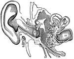
A Cross-Section of the Ear
Cross-section of the external and internal ear. a, b, and c: External ear. d: Entrance…
Dyak's ear
"Many of both sexes wear enormous ear plugs and earrings, some of which are as big around as a napkin.…
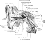
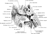
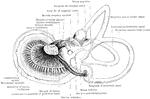
External Ear and Middle Ear
General view of the right external ear and middle. The external ear has been opened by a vertical section…
Horizontal Section of Ear
Horizontal section of right ear; upper half of section, viewed from below.
Human Ear
A diagram of the human ear. It is divided into the outer ear - A, middle ear - B, and inner ear - C.…
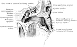
Meatus and Eustachian Tube of the Ear
Section through the external meatus, middle ear, and Eustachian tube. Labels: a, External auditory meatus;…
Middle Ear
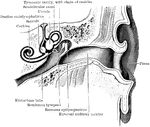
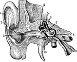
The middle ear and its bones, considerably magnified. Labels: G, the inner end of the external auditory…
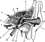
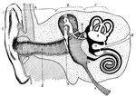
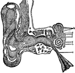
Parts of the Ear
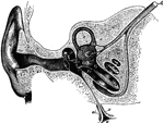
A view of all the parts of the ear, Labels: 1, The tube that leads to the internal ear. 2, The membrana…
Section of the Ear
Semidiagrammatic section through the right ear. Labels: M, concha; G, external auditory meatus; T, tympanic…
Section Through the Right Ear
Semi-diagrammatic section through the right ear. Labels: M, concha; G, external auditory meatus; T,…
Small Bones and Ligaments of the Ear
Chains of small bones and their ligaments, seen from the front in a vertical, transverse section of…
Vertical Section of Ear
Vertical transverse section of right ear; anterior half of section, viewed from behind.
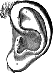
Ear-shell
"In these, which are called Ear-Shells, the animal has a shrt muzzle and two branchial plumes;…

Judas Betrays Jesus and Peter Cuts Off the Ear of Malchus
"Again therefore he asked them, Whom seek ye? And they said, Jesus of Nazareth. Jesus answered, I told…
Incus
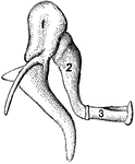
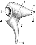
The incus, or anvil-bone. Labels: 1, body; 2, ridged articulation from the malleus; 4, processus brevis,…
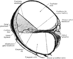
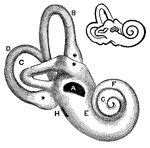
Inner Ear
1. Helix 2. Concha 3. Outer passage 4,5,6. emicircular canals 7. Oval indow 8. Cochlea 9. Eustachian…
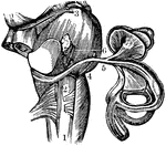
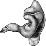
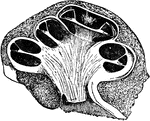
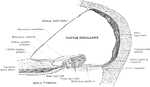
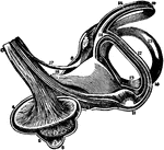
View of the Labyrinth Laid Open
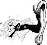
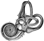
A view of the labyrinth laid open. Labels: 1, The cochlea. 2, 3, Two channels that wind two and half…
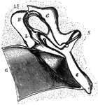
Labyrinth and Vestibule of the Ear
View, very much enlarged, of the external phase of the bony labyrinth of the left side, open, exposing…
Labyrinth of Fetus
Membranous labyrinth of a five months' fetus, viewed from its postero-mesial aspect.
Labyrinth of the Ear
View of the labyrinth in a straight position, open to show the distribution of the nerves.
Labyrinth of the Ear on the Left Side
View of labyrinth on the left side, open throughout, in order to show its structure -- enlarged.

The Labyrinth of the Inner Ear
The labyrinth of the middle ear, which is composed of a system of fluid passages.