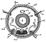
This diagram shows a transverse section of dero. c., caelom; c.l., cells of the so-called "lateral line"; d.m., dermo-muscular wall including muscles and skin; d.v., dorsal blood vessel; ect, ectoderm; ent, entoderm; g, gut; g.f., giant nerve fibers; gl, glandular cells assisting in digestion; m.c., circular muscle fibers; m.l., longitudinal muscle fibers; n, nephridium; n.v., ventral nerve chain, made up of nerve cells and nerve fibers; s, setae; v.v., ventral blood vessel.

Annelid

This diagram shows a transverse section of dero. c., caelom; c.l., cells of the so-called "lateral line";…

Diagram of a longitudinal section of an embryo, showing the different areas of the blastodermic vesicle. Labels: a, pericardium; b, bucco-pharyngeal; c, ectoderm; d, entoderm; e,placental area.

Longitudinal Section of an Embryo

Diagram of a longitudinal section of an embryo, showing the different areas of the blastodermic vesicle.…

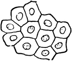
"Mosaic appearance of epithelium." — Encyclopedia Britannica, 1893

Epithelium

"Mosaic appearance of epithelium." — Encyclopedia Britannica, 1893

"Stratified epithelium." — Encyclopedia Britannica, 1893

Epithelium

"Stratified epithelium." — Encyclopedia Britannica, 1893

"A ciliated epithelium cell." — Encyclopedia Britannica, 1893

Epithelium

"A ciliated epithelium cell." — Encyclopedia Britannica, 1893

"A striated epithelium cell." — Encyclopedia Britannica, 1893

Epithelium

"A striated epithelium cell." — Encyclopedia Britannica, 1893

"A ciliated cell with one large cilium." — Encyclopedia Britannica, 1893

Epithelium

"A ciliated cell with one large cilium." — Encyclopedia Britannica, 1893

"Sense-epithelium cell of Aurelia." — Encyclopedia Britannica, 1893

Epithelium

"Sense-epithelium cell of Aurelia." — Encyclopedia Britannica, 1893

"A, blastula in which the entoderm (ent.) is produced by proliferation from ectoderm (ect.)." -Galloway, 1915

Hydroid Blastula

"A, blastula in which the entoderm (ent.) is produced by proliferation from ectoderm (ect.)." -Galloway,…
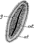
"B, ciliated planula formed by the continuance of this process. A split in the entoderm furnishes the beginning of the gastro-vascular cavity (g) of the adult." -Galloway, 1915

Hydroid Planula

"B, ciliated planula formed by the continuance of this process. A split in the entoderm furnishes the…

"Mesoderm formed by pouches from entoderm after gastrulation. A and B, early and later stages in formation of mesoderm and coelom. a, primitive gut; bp., blastopore; coe, body cavity, formed from pockets of the archenteron; ec., ectoderm; en., entoderm; m., mesoderm; m.so, body-wall mesoderm; m.sp., visceral mesoderm; s.c., segmentation cavity." -Galloway, 1915

Mesoderm Forming

"Mesoderm formed by pouches from entoderm after gastrulation. A and B, early and later stages in formation…

"Diagrammatic sagittal section of Microstomum, showing a chain of four zooids produced by fission. b, brain of the original zooid (the exponents indicating corresponding structures of the more recently formed zooids); c, ciliated pit; d, dissepiments indicating different stages in the separation of the zooids; e, eye-spot; ent, entoderm; g, gut; gl., glandular cells about the mouth; m., mouth of the original worm." -Galloway, 1915

Microstomum

"Diagrammatic sagittal section of Microstomum, showing a chain of four zooids produced by fission. b,…

Diagram of a developing ovum, seen in longitudinal section. Labels: a, pericardium; b, bucco-pharyngeal; c, ectoderm; d, entoderm; e,placental area. f, spinal cord; i, brain; g, notochord; k, extra embryonic coelom; h, dorsal wall of alimentary canal.

Developing Ovum

Diagram of a developing ovum, seen in longitudinal section. Labels: a, pericardium; b, bucco-pharyngeal;…
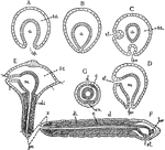
"Diagrams of stages in the metamorphosis of Polygordius, a primitive annelid. Ectoderm throughout is represented as nucleated without cell boundaries; the entoderm has the cell-boundaries shown, and the mesoderm is diagonally shaded. A, gastrula; B, same with blastopore closed; C and D represent formation of stomodaeum and proctodaeum from ectoderm; E, Trochosphere stage showing formation of segments in the posterior portion; F, adult (sagittal); G, adult (transverse). a, archenteron; bp., blostopore; br, brain; c, coelom; d, dorsal; di, dissepiments; m, mesenteron; pr., proctodaeum; s.c., segmentation cavity; st, stomodaeum; v.n., ventral nerve chain; z, zone of formation of nerve segments." -Galloway, 1915

Polygordius

"Diagrams of stages in the metamorphosis of Polygordius, a primitive annelid. Ectoderm throughout is…
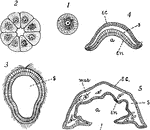
"Diagrams to illustrate the development of one of the simpler types of sponge: I, the egg; 2, section of 16- to 32-celled stage; 3, section of later stage, a ciliated larva (blastula); 4, gastrula; 5, section through older larva which has become attached by the end containing the blastopore. New openings break through by the coalescence and perforation of the ectoderm and entoderm, and and a form results... a, archenteron; bl., blastopore; ec., ectoderm; en., entoderm; mes., mesenchyma; s, segmentation cavity." -Galloway, 1915

Simple Sponge Development

"Diagrams to illustrate the development of one of the simpler types of sponge: I, the egg; 2, section…
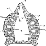
"Diagram of simple type of sponge. c, cloaca; ch, chambers, lined with flagellate entoderm; e.p., external pores; i.p., internal pores; mes., mesenchyma; o, osculum; r.c., radiating canals. In the adult sponge the canals and flagellate chambers become much more complex than figured here." — Galloway

Sponge

"Diagram of simple type of sponge. c, cloaca; ch, chambers, lined with flagellate entoderm; e.p., external…
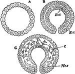
"Sections through the unilaminar (A), bilaminar (B), and trilaminar (C) conditions of the typical biastoder. Ect., ectoderm; Ent., entoderm; Mes., mesoderm; c, primitive connective tissue." — Encyclopedia Britannica, 1893

Unilaminar

"Sections through the unilaminar (A), bilaminar (B), and trilaminar (C) conditions of the typical biastoder.…