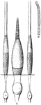
Aim
"Left eye closed, right eye looking through the notch of the rear sight so as to perceive the object…
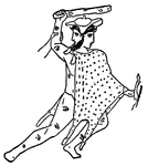
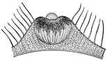
Argus
"Now Argus had a hundred eyes in his head, and never went to sleep with more than two at a time, so…
Eyes of a Bee
"The eyes, which are among the most wonderful objects in nature, are almost always of the kind called…
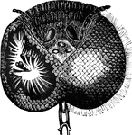
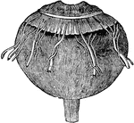
Blood Vessels of the Capsulopupillary of a Kitten
Blood vessels of the capsulopupillary membrane of a newborn kitten.
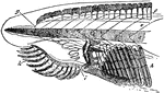
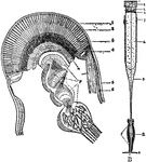
Brachiostoma
"Anterior end of body of Branchiostoma. d, chorda dorsalis; e, spinal cord; f, cartilaginous rods; g,…
Capsule of Ténon
The capsule of Ténon consists of a thin membrane which envelops the eyeball from the optic nerve…

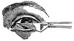
Cataract Operation
This illustration displays a way in which to correct cataracts through operation.
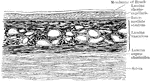
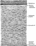
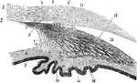
Vertical Section of the Chorioid and Sclera
Vertical section of the chorioid and inner part of the sclera.
Cones and Rods of Retina
A. A cone and two rods from the human retina (modified from Max Schultze); B. Outer part of rod separated…
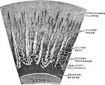
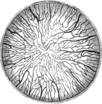
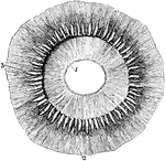
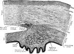
Magnified Corona Ciliaris
A portion of the corona ciliaris magnified. The ciliary processes and the ciliary folds.
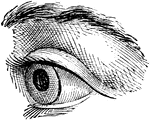


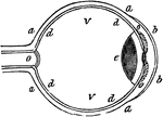
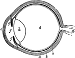
Eye
"Next in order is the aqueous humor, b, e, in the middle of which is the iris, d, c. Behind the pupil…
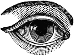
Eye
"a, sclerotic membrane; b, cornea; d, retina; o, optic nerve; v, vitreous humor." -Comstock 1850
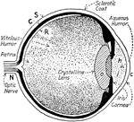
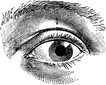
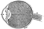
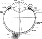
Eye
The human eye. Labels: a, crystalline lens; b, retina; c, cornea; d, sclerotic; e, choroid; g, ciliary…
The Eye and its Muscles
The eye and its muscles. Labels: o, the nerve of sight; a, one of the muscles of the eye.

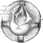
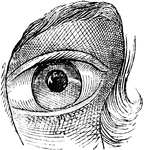
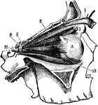
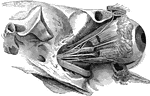
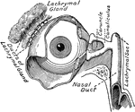
Eye and Lachrymal Gland
Front view of left eye, with eyelid partly removed to show lachrymal gland (tear-producing gland), and…
Ciliary Processes of the Eye as Seen from Behind
Ciliary processes, as seen from behind. Labels: 1, posterior surface of the iris, with the sphincter…
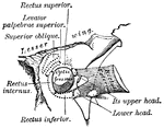
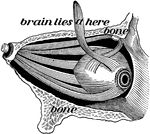
Eye Muscles
"The external bones of the temple are supposed to be removed in order to render visible the muscular…
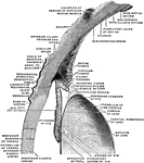
Eye Nerves of a Horse
Right orbit opened to show the nerves of the eye. Labels: a, optic; b, motor oculi; c, pathetic; d,…
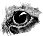
Eye of a Crayfish
A, section through the compound eye of a crayfish. Labels: 1, cornea; 2, crystaline cones; 3, retinulae;…
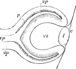
Eye of a Lizard
Longitudinal section through the pineal eye of a lizard. The eye is located in the middle of the dorsal…
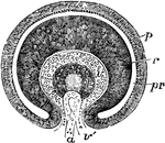
Eye of Fetus of Four Weeks
Diagrammatic sketch of a vertical longitudinal section through the eyeball of a human fetus of four…
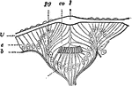
Eye of Fetus of Four Weeks
Transverse vertical section of the eyeball of a human embryo of four weeks. The anterior half of the…
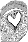
Eye of Gasteropod
Section through the cup-shaped eye of a gastropod. Labels: e, epithelium covering body; cv, vitreous…
Eye of Waterbeetle
Section through the eye of a waterbeetle. Labels: l, chitinous lens; cv, transparent cells; pg, pigment…
Optical Position and Size of Image Through Lens in Front of Eye
The illustration of putting lenses in front of the eye. The focal point of the image is reflected into…
A Section of the Eye Seen from Within
A section of the eye seen from within. Labels: 1, The divided edge of the three coats. 2, The pupil.…
A Section of the Eye
A section of the eye. Labels: 1, The sclerotic coat. 2, The cornea. 3, The choroid coat. 6, The iris.…

Astor to Philip's Right Eye
"It was during the siege of Methone that Philip had the misfortune to lose on of his eyes. A random…
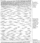
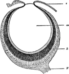
Choroid Coat of the Eye
The eye, after cutting away the sclerotic coat and cornea, to show the vessels of the choroid coat;…
Ciliary Processes of the Eye
Section through the eye carried through the ciliary processes. Labels: 1, Cornea; 2, membrane of Descemet;…
Cornea too Concave on Eye
"...and the cornea will become too flat, or not suffciently convex, to make the rays of light meet at…
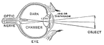
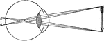
Diagram of the Eye
"Diagram illustrating the Manner in which the Image of an Object is inverted on the Retina." — Blaisedell,…
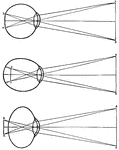
Human Eye
Diagrams of how an image is displayed with a normal eye (top image), myopic or nearsighted eye (middle…

Lens of the Eye
Lens of the eye. The rays of light are brought nearer together by the lenses of the eye, just as they…
Lens of the eye
"Diagram showing the Change in the Lens during Accomadation. On the right the lens is arranged for distant…
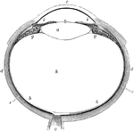
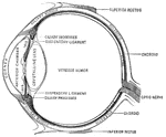
Median Vertical Anteroposterior Section of Eye
"Human Eye, in Median Vertical Anteroposterior Section. (Ciliary processes shown, through not all lying…

Eye Focusing on Object
"Showing how the image of an object which is seen is formed on the retina of the eye." —Croft 1917
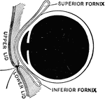
Sagittal Section of the Eye
Sagittal section of the eye, showing superior and inferior fornices of the conjuctiva.
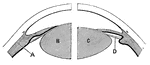
Sagittal Section Through the Eye
The upper half of a sagittal section through the front of the eyeball.
Section of the Eye
Section of the eye, showing the relations of the cornea, sclera, and iris, together with the Ciliary…
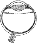
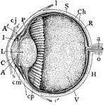
The Eye
The eye. Labels: a, sclerotica; e, cornea; b, choroid; d, optic nerve; f, aqueous humor; g g , iris;…