"Section through the left eye, closed. 1, lifting muscle; 2, upper straight muscle; 3, optic nerve; 4, fatty cushion; 5, lower straight muscle; 6, vitreous humor; 7, lower cross muscle; 8, lower eyelid; 9, upper eyelid; 10, crystalline lens." -Foster, 1921

Eye Section

"Section through the left eye, closed. 1, lifting muscle; 2, upper straight muscle; 3, optic nerve;…
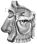
"The Relative Position of the Lachrymal Apparatus, the Eyeball, and the Eyelids. <I>A</em>, lachrymal canals, with the minute orifices represented as two black dots to the right; <em>B</em>, tendon attached to a muscle which surrounds the circumference of the orbit and eyelids; under <em>B</em> is seen the lachrymal sac. The minute openings of the Meibomian glands are seen on the free margins of the eyelids. Below <em>A</em> is seen a small conical elevation, with black dots (the lachrymal papilla, or caruncle)." &mdash; Blaisedell, 1904

Eyeball

"The Relative Position of the Lachrymal Apparatus, the Eyeball, and the Eyelids. A, lachrymal canals,…

"<em>A</em>, attachment of tendon connected with the four recti muscles; <em>B</em>, external rectus, divided and turned downward, to expose the internal rectus; <em>C</em>, inferior rectus; <em>D</em>, internal rectus; <em>E</em>, superior rectus; <em>F</em>, superior oblique; <em>H</em>, pulley and reflected portion of the superior oblique; <em>K</em>, inferior oblique; <em>L</em> and <em>M</em>, portions of the muscle which raises the upper eyelid; to the right of <em>D</em> and to the left on the same line are seen cut ends of the optic nerve." &mdash; Blaisedell, 1904

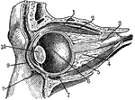
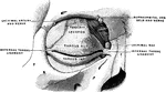
Muscles of the eyeball

"A, attachment of tendon connected with the four recti muscles; B, external rectus,…

Side view of the eyeball. Labels: a, the eyeball, and b,b, are the upper and lower sides. Now in order to prevent dust and other bodies from working their way between the ball and the lid, through passages at c,c, into the socket of the eye where they would excite great inconvenience and pain, we find the common skin of the eye lids d,d, after covering their edges turn in a little way between the lid and the ball, and then turn back and are reflected over the surface of the cornea; where, to prevent the obstruction of vision, it becomes perfectly transparent.

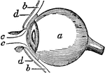
Side View of the Eyeball

Side view of the eyeball. Labels: a, the eyeball, and b,b, are the upper and lower sides. Now in order…
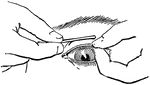
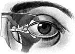
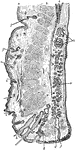
A vertical section of the upper eyelid.

Eyelid

A vertical section of the upper eyelid.

The tarsi and their ligaments.

Eyelid

The tarsi and their ligaments.

"Showing how the upper eyelid may be everted with a pencil or penholder." — Blaisedell, 1904

Everted eyelid

"Showing how the upper eyelid may be everted with a pencil or penholder." — Blaisedell, 1904

The Meibomian glands, ect., seen from the inner surface of the eyelids.

Inner Surface of the Eyelid

The Meibomian glands, ect., seen from the inner surface of the eyelids.

Vertical section through the upper eyelid. Labels: a, Skin; b, Orbicularis palpebrarum; b', Marginal fasciculus of Orbicularis (ciliary bundle); c, Levator palpebrae; d, Conjuctiva; e, Tarsal plate; f, Meibomian gland; g, Sebaceous gland; h, Eyelashes; i, Small hairs of skin; j, Sweat glands; k, Accessory lacrimal glands.

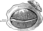
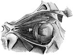
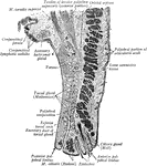
Vertical Section Through Eyelid

Vertical section through the upper eyelid. Labels: a, Skin; b, Orbicularis palpebrarum; b', Marginal…

<em>A</em> and <em>C</em>, lachrymal canals; <em>B</em>, lachrymal sac; <em>D</em>, small muscle which serves to compress the lachrymal sac; <em>F</em>, glands upon the inner surface of the eyelids, with ducts opening upon the free margins of the eyelids; <em>H</em>, great openings, or antrum, of the upper jawbone. The oil glands of the nose are plainly shown." &mdash; Blaisedell, 1904

Eyelids and lachrymal apparatus

A and C, lachrymal canals; B, lachrymal sac; D, small muscle which…