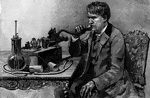
Differential Lens
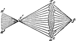
"Differential Lens.—Horizontal divergence may be obtained to any required amount by varying the radius…
Differential Lens
"Differential Lens.—Horizontal divergence may be obtained to any required amount by varying the radius…
Differential Lens
"Differential Lens.—Horizontal divergence may be obtained to any required amount by varying the radius…
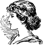
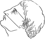
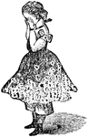
Hat Display
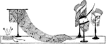
This is an early 20th century hat display. It includes three hats, a long shawl, purse, and hat pins…
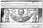
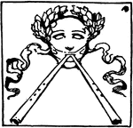
Encarpus
"In architecture, a sculptured ornament in imitation of a garland or festoon of fruits, leaves, or flowers,…
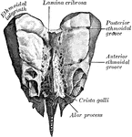
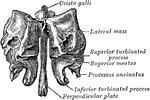
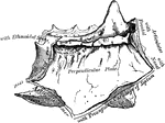
Perpendicular Plate of Ethmoid
Perpendicular plate of ethmoid, shown by removing the right lateral mass.
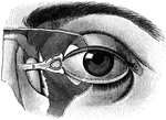
Eyeball
"The Relative Position of the Lachrymal Apparatus, the Eyeball, and the Eyelids. A, lachrymal canals,…
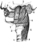
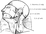
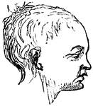
Face
A side view of face. 1 and 2: Trachea. 3: Esophagus. 4, 5, and 6: Muscles. 7: Submaxillary. 8: Parotid…
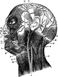
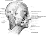
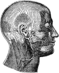
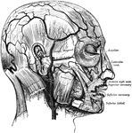
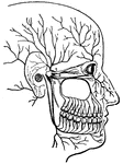
Arteries of the Face and Head
The arteries of the face and head. Labels: 1, common carotid; 2, internal carotid; 3, external carotid;…
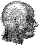
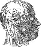
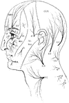
Nerves of the Face and Scalp
Nerves of the face and scalp. Labels: 1, attrahensaurem; 2, anterior belly occipito-frontalis; 3, auriculo-temporal…
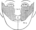
Development of the Face
Showing the development of the face. F.N.P., Part formed from the frontonasal process; L, from its lateral…
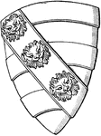
Heraldic Shield with a Lion's Face
The shield of a knight of the time of Edward II. The lion's face is a common charge in heraldry.
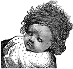
Mesial Sagittal Section of Child's Face
Anterior portion of mesial sagittal section of child's head, probably of about three year.
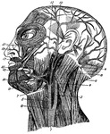
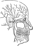
Facial Arteries
The arteries of the face and scalp. The muscle tissue of the lips must be supposed to have been cut…

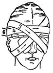
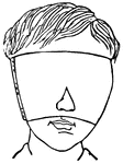
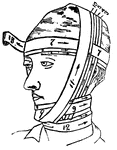
Facial Bandage
This illustration shows a method of applying a bandage to the face to cover both eyes.
Facial Muscles
Muscles of the face, jaw and neck. 1, longus colli; 2, rapezius; 3, sterno-hyoid; 4, sterno-mastoid;…
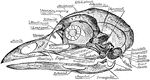
Skull of a Common Fowl
"Fig. 62 Skull of common fowl, enlarged. from nature by Dr. R.W. Shufeldt, U.S.A. The names of bones…


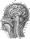
Sagittal Section of the Head and Neck
Sagittal median section of the head and neck. The head is thrown backward into complete extension which…
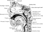
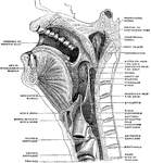
Section of the Head and Neck
Section of head and neck from front to back. Labels: 1, windpipe; 2, larynx; 3, spinal marrow; 4, pharynx;…
Sensory Nerves to the Head and Neck
Distribution of sensory nerves to the head and neck. Labels: Ophth, ophthalmic division of the fifth…
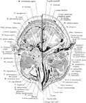
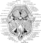
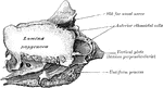
Cross Section of Head Exposing Maxillary Sinus
Section of the head immediately below the orbits, at the level of Reid's base line exposing the maxillary…