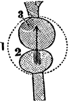
A Diagram of the Heart of a Fish
A diagram of the heart of a fish. Labels: 1, Pericardium. 2, The ventricle that receives the blood from…
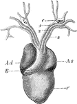
Heart of a Frog
The heart of a frog (Rana esculenta) from the front. Labels: V, ventricle, Ad, right auricle; As, left…
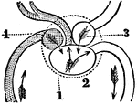
A Diagram of the Heart of a Reptile
A diagram of the heart of a reptile. Labels: 1, Pericardium. 2, Single ventricle. 3, Left auricle. 4,…
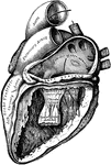
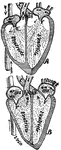
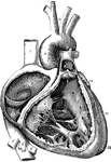
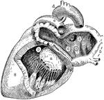
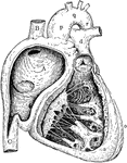
Heart with Left Auricle and Ventricle Laid Open
The left auricle and ventricle laid open, the posterior walls of both being removed.
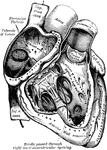
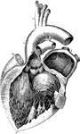
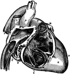
Heart with Right Auricle and Ventricle Laid Open
The right auricle and ventricle laid open, the anterior walls of both being removed.
A Diagram of the Heart
A diagram of the heart. Labels: 1, Right and left auricle. 2, Right and left ventricle. 3, 4, The pericardium.…
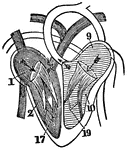
A Diagram of the Heart
A diagram of the heart. Labels: 1, Right auricle. 2, Right ventricle. 9, Left auricle. 10, Left ventricle.…
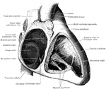
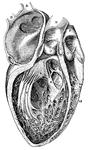
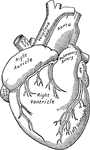
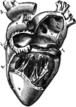
Anatomy of Heart
A complex anatomical view of the heart. RA is the right atrium, or auricle, which receives the deoxygenated…
Anterior View of the Heart
Anterior view of the heart, dissected, after long boiling to show the superficial muscular fibers. The…
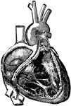
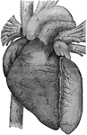
Anterior View of the Heart
An anterior view of the heart in a vertical position with its vessels injected.

Auricle and Ventricle of the Heart
The cavities of the right auricle and right ventricle of the heart.
Beating heart
"Diagram of the rush of blood when the heart beats. The valves v open above are closed below while the…
Cavities of the heart
"A, B, right pulmonary veins, S, openings of the left pulmonary veins; E, D, C,…
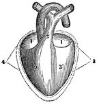
Chambers of the Heart
The chambers of the heart. Labels: A, right ventricle; B, left ventricle; C, right auricle; D, left…
Heart, Compartments of
Diagram showing the compartments of the heart. The smaller compartments are the auricles (also known…
A Diagram of the Heart
The right auricle and ventricle opened, and a part of their right and anterior walls removed, so as…
A Diagram of the Heart
The left auricle and ventricle opened and a part of their anterior and left walls removed. The pulmonary…
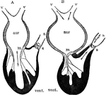
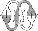
Diagram of the Action of the Heart
Diagram to illustrate the action of the heart. Labels: aur, auricle (atrium); vent. ventricle; v, veins;…
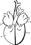
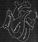
Diagram of the heart
"Diagram of the passages of the heart. 1. left auricle. 2. left ventricle, 3. Right auricle. 4. Right…
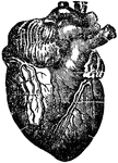
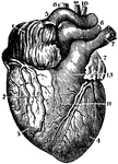
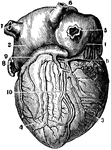
Heart, Front View of
A representation of the heart as it really appears showing the front view. At a is the right…
Interior of the Heart
Diagram of the interior of the heart. Labels: A, aorta; PA, pulmonary artery; VCI and VCS, vena cava…
Left Side of Heart
Left side of heart. Labels: 1, cavity of left auricle (atrium); 3, opening of right pulmonary veins;…
Left Side of the Heart
Left side of the heart, showing the left auricle (atrium) and left ventricle).
Posterior View of the Heart
A posterior view of the heart in a vertical position and with its vessels injected.
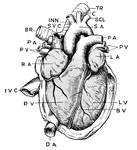
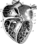
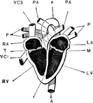
Right Side of Heart
Right side of heart. Labels: A, cavity of right ventricle; B, superior vena cava; C, inferior vena cava;…
Right Side of the Heart
Right side of the heart, showing the right auricle (atrium) and right ventricle).
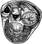
Section of Heart Showing Valves
Section of heart at level of valves. Labels: P, pulmonary artery, with flaps of semilunar valve open;…
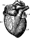
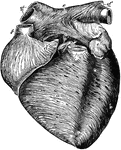
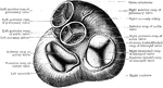
The Heart
The heart. Labels: RA, right auricle, RV; right ventricle; LA, left auricle; LV, left ventricle.
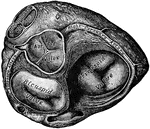
Ventricle of the Heart
The bases of the ventricle of the heart, showing the auriculoventricular, aortic, and pulmonary orifices…
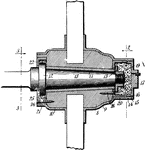
Wheel Hub
The central part of a car wheel (or fan or propeller etc) through which the shaft or axle passes
Intestinal absorption
"A, a fold of peritoneum; B, lacteals and lymphatic glands; C, veins of intestines;…
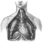
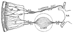
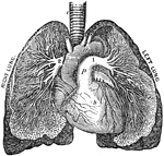
Lungs
"Relative Postion of the Lungs, the Heart, and Some of the Great Vessels belonging to the latter. A,…
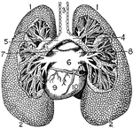
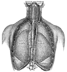
Lungs
"The Lungs. 1, Summit of lungs. 2, Base of lungs. 3, Trachea. 4, Right bronchus. 5, Left bronchus. 6,…
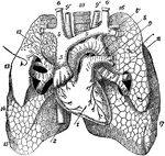
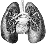
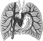
Anterior View of the Lungs and Heart
Anterior view of the lungs and heart. Labels: 1, heart; 2, inferior vena cava; 3, superior vena cava;…
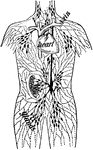
Lymph Vessels
"The lymph vessels of the body. rc, the thoracic duct; lac, the lacteals taking the lymph and fatty…
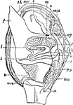
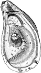
Mollusc Anatomy
"Anatomy of an Acephalous Mollusc (Mactra): s, stomach; ii, intestine; ag, anterior ganglions; pg, posterior…
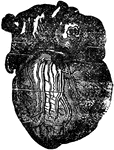
Heart Muscle
The muscular fibres of the heart, unlike those of most involuntary muscles, are striated.
Muscular Fiber Cells from the Heart
Muscular fiber cells from the heart. The fibers which lie side by side are united at frequent intervals…
Oyster
"Anatomy of the Oyster. A. Hinge or anterior umbonal end of the left valve of an adult oyster, upon…
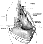
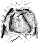
Pericardium Ligaments
The pericardium is a conical serofibrous sac in which the hear and the commencement of the great vessels…
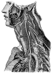
Trunk of the Pneumogastric Nerve
"Showing its distribution by its branches and ganglia to the larynx, pharynx, heart, lungs, and other…
Poetic Rebus
"I long to lay this aching head and wounded heart beneath the soil, To slumber in that dreamless bed…
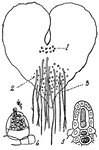
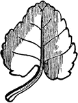
Prothallus (under side)
The flat, heart shaped body which results from the development of a spore of a fern.
The Pulmonary Artery
The pulmonary artery. Labels: t, The trachea. h, The heart. a, The aorta. p, The pulmonary artery. 1,…
A Diagram of Pulmonary Circulation
A diagram of pulmonary circulation. Labels: 1, Descending vena cava. 2, Ascending cava vein. 3, Chamber…
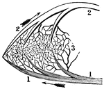
A Portion of the Pulmonic Circulation
A portion of the pulmonic circulation. 1, A branch of the artery that carries the impure blood to the…