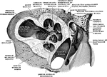
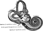
The cochlea laid open.

Cochlea Laid Open

The cochlea laid open.

Vertical section through the right cochlea, medial portion, viewed from the lateral side.

Vertical Section Through the Cochlea

Vertical section through the right cochlea, medial portion, viewed from the lateral side.
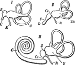
Internal ear of different vertebrates. I, fish; II, bird; III, mammal. Labels: U, utriculus with semicircular canals; S, sacculus; US, utriculus and sacculus; C, cochlear duct; L, lagena; Cr, canalis reuniens; R, recessus vestibuli.

Internal Ear of Vertebrates

Internal ear of different vertebrates. I, fish; II, bird; III, mammal. Labels: U, utriculus with semicircular…
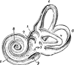
View of the interior of the left labyrinth. The bony wall of the labyrinth is removed superiorly and externally. Labels: 1, fovea hemielliptica; 2m fovea hemispherica; 3, common opening of the superior and posterior semicircular canals; 4, opening of the aqueduct of the vestibule; 5, the superior, 6, the posterior, and 7, the external semicircular canals; 8, spiral tube of the cochlea (scala tympani); 9, opening of the aqueduct of the cochlea; 10, placed on the lamina spiralis in the scala vestibuli.

Interior of the Left Labyrinth

View of the interior of the left labyrinth. The bony wall of the labyrinth is removed superiorly and…
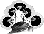
The osseous labyrinth laid open.

Osseous Labyrinth

The osseous labyrinth laid open.

The osseous labyrinth in vertical section. The broken, white lines indicate the position of the basilar membrane of the canal of the cochlea.

Osseous Labyrinth in Vertical Section

The osseous labyrinth in vertical section. The broken, white lines indicate the position of the basilar…

Diagram to illustrate the action of the ossicles of the middle ear in the conduction of sound to the internal ear.

Ossicles of the Middle Ear

Diagram to illustrate the action of the ossicles of the middle ear in the conduction of sound to the…