Clipart tagged: ‘knee’
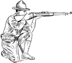
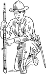
Aim kneeling
"In aiming kneeling, the left elbow rests on the left knee, point of elbow i nfront of kneecap." —…
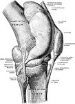
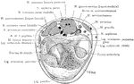
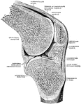
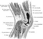
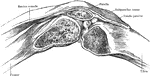
Dissection of Knee Joint From Front
Dissection of knee joint from the front with patella thrown down.
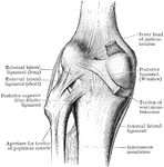
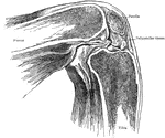
Knee Joint from Lateral Surface
Right knee joint from the lateral surface. The joint cavity and several bursae have been injected with…

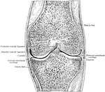
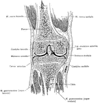
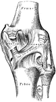
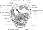
Frontal Section Through Knee Joint
Frontal section through middle of right knee joint. Seen from behind.
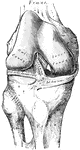
Frontal Section Through Knee Joint
Frontal section through knee joint, showing articulating surfaces and epiphyseal lines.

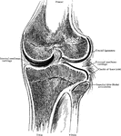
Sagittal Section Through Knee Joint
Right knee joint. Sagittal section through the external condyle of the femur. Mesal half of section,…
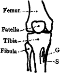
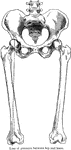
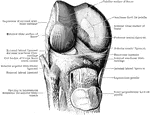
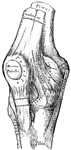
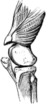
Bones of the Knee
Bones of the knee also showing muscle. Labels: s, insertion of the sartorius; g, insertion of the gracilis.
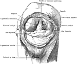
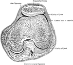
Patella Removed from Knee
Patella removed from right knee, which is strongly flexed to show alar ligaments and ligamentum mucosum.…
Sagittal Section Through Knee
Sagittal section of the right knee, viewed from the outer side. The joint cavity proper lies to each…
Muscles of the leg
"Muscles of the leg showing how they pass into tendons at the ankle." —Davison, 1910
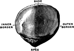
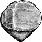
Patella
View of the posterior surface of the right patella, showing diagrammatically the areas of contact with…
Position of Patella in Relation to Condyles of Femur with Knee Flexed
Showing position of patella in relation to condyles of femur with knee flexed at a right angle.
Position of Patella in Relation to Condyles of Femur with Knee Partially Flexed
Showing position of patella in relation to condyles of femur with knee partially flexed.
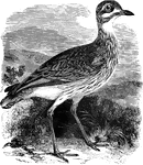
Great Plover
The great plover, also known as the thick-knee averages about seventeen inches in length and ranges…
Thigh Muscles
"Some of the Larger Muscles on the back of the Thigh. Powerful tendons at the hip and on the back of…