Clipart tagged: ‘labyrinth’
Ear
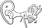
Interior of the ear. There is external to the head a wide-mouthed tube, or ear-trumpet (a), for catching…
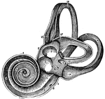
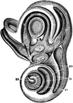
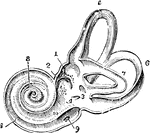
View of the Labyrinth Laid Open
A view of the labyrinth laid open. Labels: 1, The cochlea. 2, 3, Two channels that wind two and half…
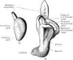
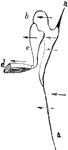
Labyrinth of Fetus
Membranous labyrinth of a five months' fetus, viewed from its postero-mesial aspect.
Labyrinth of the Ear
View of the labyrinth in a straight position, open to show the distribution of the nerves.
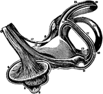
Labyrinth of the Ear on the Left Side
View of labyrinth on the left side, open throughout, in order to show its structure -- enlarged.

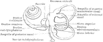
The Labyrinth of the Inner Ear
The labyrinth of the middle ear, which is composed of a system of fluid passages.
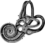
The Labyrinth of the Inner Ear
A view of the labyrinth of the left side laid open in its whole extend, so as to show its structure…
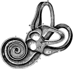
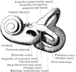
Bony Labyrinth of the Inner Ear
A highly magnified view of the external face of the bony labyrinth of the left side, opened so as to…
Development of Labyrinth
A, Left labyrinth of a human embryo of about four weeks; B, left labyrinth of a human embryo of about…
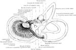
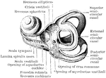
Interior of the Left Labyrinth
View of the interior of the left labyrinth. The bony wall of the labyrinth is removed superiorly and…

Medal of Crete
A medal of Crete, representing a Minotaur and the labyrinth in which he was confined.
Ossicles of the Middle Ear
Diagram to illustrate the action of the ossicles of the middle ear in the conduction of sound to the…