Clipart tagged: ‘Lamella’
Section of Bone
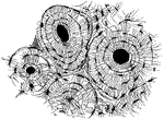
A small piece of bone, ground very thin and highly magnified. "If a bit of bone is still more magnified…
Lamella of Kitten's Cornea
Surface view of part lamella of kitten's cornea, prepared first with caustic potash and then with nitrate…
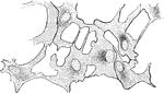
Section of Rabbit's Cornea
Vertical section of rabbit's cornea, stained with gold chloride. Labels: e, Laminated anterior epithelium.…
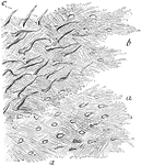
Lamellae
Lamellae torn off from a decalcified human parietal bone at some depth from the surface. Labels: a,…
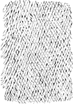
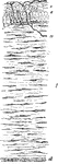
Reticular Structure of a Lamellae
Shown is a thin layer peeled off from a soft bone, which is intended to represent the reticular structure…
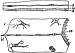
Medusae
"Portions of sections through the disc of medusae. The upper one of Lizzia, the lower of Aurelia. el,…