Clipart tagged: ‘lungs’
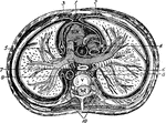
Bird Arteries
Arteries of the trunk of a bird. 1: The aorta. 2: The vena cava. 3: A cerebral artery. The small lines…
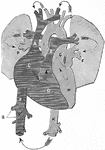
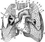
Branchi and Blood Vessels
Branchi of the lungs, the heart, and blood vessels. Labels: 1, left auricle; 2, right auricle; 3, left…
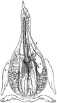
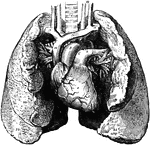
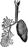
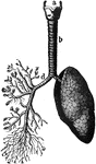
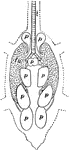
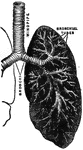
Bronchia and Veins of the Lungs
View of the bronchia and veins of the lungs, exposed by dissection, as well as the relative position…
Circulation of Blood
A diagram illustrating the circulation of the blood. Labels: A, vena cava descending (superior); Z,…
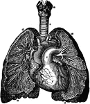
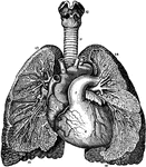
Heart and Lungs
The heart and lungs. 1, right ventricle; 3, right auricle (atrium); 6, 7, pulmonary artery; 9, aorta;…
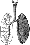
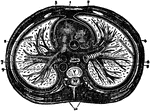
Relative Position of the Heart and Lungs
A view of the bronchia and blood vessels of the lungs, as shown by dissection, as well as the relative…
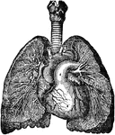
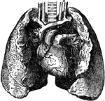
Heart and Lungs
The heart, showing its relative position to the lungs. The heart is almost wholly covered up by the…
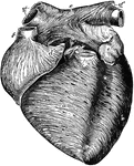
Anterior View of the Heart
Anterior view of the heart, dissected, after long boiling to show the superficial muscular fibers. The…
Lungs
The lung is the essential organ of respiration in air-breathing vertebrates. Its principal function…
Lungs
"The Lungs. 1, Summit of lungs. 2, Base of lungs. 3, Trachea. 4, Right bronchus. 5, Left bronchus. 6,…
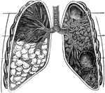
Lungs and Air Passages
The lungs and air passages seen from the front. On the left of the figure the pulmonary tissue has been…
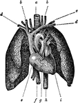
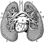
Anterior View of the Lungs and Heart
Anterior view of the lungs and heart. Labels: 1, heart; 2, inferior vena cava; 3, superior vena cava;…
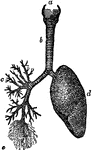
Structure of the Lungs
Diagram showing the structure of the lungs. At d is the left lung, and at c are represented…
The Lungs
The lung, which are the two essential organs of respiration contained in the cavity of the thorax.
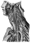
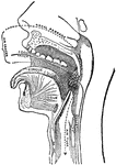
Trunk of the Pneumogastric Nerve
"Showing its distribution by its branches and ganglia to the larynx, pharynx, heart, lungs, and other…
Respiratory Mechanism
"The respiratory mechanism consists of the lungs, a series of minute air chambers with a network of…
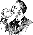
Sick Man Sneezing
A sneeze is a semi autonomous, convulsive explosion of air form the lungs through the nose and mouth,…
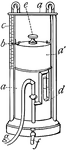
Spirometer
"A contrivance for measuring the extreme differential capacity of the human lungs. The instrument most…

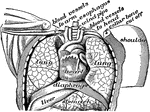
Thorax and Abdomen
"Thorax and abdomen. 1, 1, 1, 1. Muscles of the chest. 2, 2, 2, 2. Ribs. 3, 3, 3. Upper, middle and…
The Thorax
Diagram of a transverse section of the thorax. Labels: 1, anterior mediastinum; 2, internal mammary…
Transverse Section of the Thorax
Transverse section of the thorax. Labels: 1, anterior mediastinum; 2, internal mammary vessels; 3, triangularis…
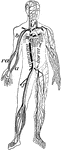
Veins and arteries
"Chief veins and arteries of the body. a, place of the heart; the veins are in black. On the right side…
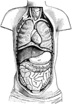
Ventral Cavity of the Body
Diagram of the Body opened from the front to show the contents of the ventral cavity. Labels: d, diaphragm;…