Alimentary canal of a bird. Labels: a, ingluvies; b, proventriculus; c, pancreas; d, duodenum; e, liver; f, biliary cyst and duct; g, pancreatic duct; h, intestice; i, the to caeca; k, rectum; l, right ureter; m, oviduct; n, its aperture in cloaca; o, opening of cloaca.

Alimentary Canal of a Bird

Alimentary canal of a bird. Labels: a, ingluvies; b, proventriculus; c, pancreas; d, duodenum; e, liver;…
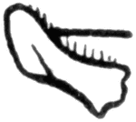
The mandible of the honey-bee.

Honey Bee

The mandible of the honey-bee.

A beetle named after its large, distinctive mandibles found on the males.

Stag Beetle

A beetle named after its large, distinctive mandibles found on the males.

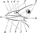
"Fig. 26 - Parts of a Bill. a, side of upper mandible; b, culmen; c, nasal fossa; d, nostril; e(see below); f, gape, or whole commissural line; g, rictus; h, commissural point or angle of the mouth; i, ramus of under jaw; j, tomia of under mandible (the reference lines e should have been drawn to indicate the correct tomia of upper mandible):k, angle of gonys; l, gonys; m, side of under mandible; n, tips of mandibles." Elliot Coues, 1884

The Parts of a Bird Bill

"Fig. 26 - Parts of a Bill. a, side of upper mandible; b, culmen; c, nasal fossa; d, nostril; e(see…
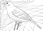
"fig. 25 - Topography of a Bird. 1, forehead (frons). 2, lore. 3, circumocular region. 4, crown (vertex). 5, eye. 6, hind head (occiput). 7, nape (nucha). 8, hind neck (cervix). 9, side of neck. 10, interscapular region. 11, dorsum, or back proper, including 10. 12, notaeum, or upper part of body proper, including 10, 11, and 13. 13, rump (uropygium). 14, upper tail-coverts. 15, tail. 16, under tail-coverts (crissum). 17, tarsus. 18, abdomen. 19, hind toe (hallux). 20, gastraeum, including 18 and 24. 21, outer and fourth toe. 22, middle and third toe. 23, side of the body. 24, breast (pectus). 25, primaries. 26, secondaries. 27, tertiaries; nos. 25, 26, 27 are all coverts. 28, primary coverts. 29, alula, or bastard wing. 30, greater coverts. 31, median coverts. 32, lesser coverts. 33, the "throat," including 34, 37, 38. 34, jugulum or lower throat. 35, auriculars. 36, malar region. 37, gula, or middle throat. 38, mentum, or chin. 39, angle of commissure, or corner of mouth. 40, ramus of under mandible. 41, side of under mandible. 42, gonys. 43, apex, or tip of bill. 44, tomia, or cutting edges of the bill. 45, culmen, or ridge of upper mandible, corresponding to gonys. 46, side of upper mandible. 47, nostril. 48, passes across the bill a little in front of it base." Elliot Coues, 1884

Topography of a Bird

"fig. 25 - Topography of a Bird. 1, forehead (frons). 2, lore. 3, circumocular region. 4, crown (vertex).…
An illustration of a cladognathus cinnamomeus beetle.

Cladognathus Cinnamomeus

An illustration of a cladognathus cinnamomeus beetle.

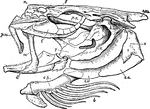
"The skull of a cod. b, branchiostegal rays born on c.h., the ceratohyal bone; d, dentary portion of the mandible; f, frontal; h.m., hyomandibular; i.o., interoperculum; l, lachrymal; m, maxilla; n, nasal; o, operculum; p.m., premaxilla; p.o., preoperculum; p.s., parasphenoid; q, quadrate; s.o., sub-operculum; s.oc., supra-occipital." — Galloway

Cod Skull

"The skull of a cod. b, branchiostegal rays born on c.h., the ceratohyal bone; d, dentary portion of…
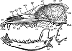
"Longitudinal and Vertical section of the skull of a dog, with mandible and hyoid arch. an, anterior narial aperture; MT, maxillo-turbinal bone; ET, the ethmo-turbinal; Fr, frontal; Pa, pariental; IP, interparietal; SO, supra-occipital; ExO, sphenoid; OS, orbito-sphenoid; PS, presphenoid; Pl, palatine; Vo, vomer; Mx, maxilla; PMx, premaxilla; sh, stylo-hyal; eh, epi-hyal; ch, cerato-hyal; bh, basihyal; th, thyro-hyal; s, symphysis of mandible; cp, coronoid process; cd, condyle; a, angle; id, inferior dental canal. The mandible is displaced downwards, to show its entire form; the * indicated the part of the cranium to which the condyle is articulated." —The Encyclopedia Britannica, 1903

Dog Skull

"Longitudinal and Vertical section of the skull of a dog, with mandible and hyoid arch. an, anterior…
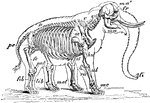
"Skeleton and Outline of African Elephant (Elephas or Loxodon africanus). fr, frontal; ma, mandible; ma', malar; fi, "finger" at end of trunk; C, cervical vertebrae; D, dorsal vertebrae; D, dorsal vertebrae; pe, pelvis; sc, scapula; st, sternum; hu, humerus; ul, ulna; ra, radius; mc, metacarpus; fe, femur; pat, patella; tib, tibia; fib, fibula; met, metatarsus." -Whitney, 1911

African Elephant Skeleton

"Skeleton and Outline of African Elephant (Elephas or Loxodon africanus). fr, frontal; ma, mandible;…

"Fig. 62 Skull of common fowl, enlarged. from nature by Dr. R.W. Shufeldt, U.S.A. The names of bones and some other parts are printed, requiring no explanation; but observe the following points: The distinction of none of the bones composing the brain-case (the upper back expanded part) can be found in a mature skull. The brain is contained between the occipital, sphenoidals, squamosals, parietals and part of frontal; the ethmoidals belong to the same group of cranial bones proper. All other bones, excepting the three otic ear-bones, are bones of the face and jaws. The lower jaw, of five bones, is drawn detached; it articulates by the black surface marked articular with the prominence just above- the quadratic bone. Observe that from this quadrate a series of bones quadrato-jugal, jugal, maxillary-makes a slender rod running to the premaxillary; this is the zygoma, or jugal bar. Observe from the quadrate also another series, composed of pterygoid and palatine bones, to the premaxillary; this is the pterygo-palatine bar; it slides along a median fixed axis of the skull, the rostrum, which bears the loose vomer at its end. The under mandible, quadrate, pterygoid, and vomer are the only movable bones of this skull. But when the quadrate rocks back and forth, as it does by its upper joint, its lower end pulls and pushes upon the upper mandible, by means of the jugal and pterygo-palatine bars, setting the whole scaffolding of the upper jaw in motion. This motion hinges upon the elasticity of the bones of the forehead, at the thin place just where the reference-lines from the words "lacrymal" and "mesethmoid" cross each other. The dark oval space behind the quadrate is the external orifice of the ear; the parts in it to which the three reference-lines go are diagrammatic, not actual representations; thus, the quadrate articulates with a large pro-otic as well as with the squamosal. The great excavation at the middle of the figure, containing the cirlet of the unshaded bones, is the left orbital cavity, orbit, or socket of the eye. The mesethmoid includes most of the background of this cavity, shaded diagonally. The upper one of the two processes of bone extending into it from behind is post-frontal or sphenotic process; the under one (just over the quadrate) is the squamosal process. A bone not shown, the presphenoid, lies just in front of the oval black space over the end of basisphenoid. This black oval is the optic foramen, through which the nerve of sight passes from the brain-cavity to the eye. The black dot a little behind the optic foramen is the orifice of exit of a part of the trifacial nerve. The black mark under the letters "on" of the word "frontal" is the olfactory foramen, where the nerve of smell emerges from the brain-box to go to the nose. The nasal cavity is the black space behind nasal and covered by that bone, and in the oval blank before it. The parts of the beak covered by horn are only premaxillary, nasal, and dentary. The condyle articulates with the first cervical vertebra; just above it, not shown, is the foramen magnum, or great hole through which the spinal medulla, or main nervous cord, passes from the spinal column. The basioccipital is hidden, excepting its condyle; so is much of the basisphenoid. The prolongation forward of the basisphenoid, marked "rostrum," and bearing the vomer at its end, is the parasphenoid, as far as its thickened under border is concerned. Between the fore end of the pterygoid and the basisphenoidal rostrum, is the site of the basipterygoid process, by which the bones concerned articulate by smooth facets; further forward, the palatines ride freely upon the parasphenoidal rostrum. In any passerine bird , the vomer would be thick in front, and forked behind, riding like the palatine upon the rostrum. The palatine seems to run into the maxillary in this view; but it continues on to premaxillary. The maxillo-palatine is an important bone which cannot be seen in the figure because it extends horizontally into the paper from the maxillary about where the reference life "maxillary" goes to that bone. The general line from the condyle to the end of the vomer is the cranial axis, basis cranii, or base of the cranium. This skull is widest across the post-frontal; next most so across the bulge of the jugal bar." Elliot Coues, 1884

Skull of a Common Fowl

"Fig. 62 Skull of common fowl, enlarged. from nature by Dr. R.W. Shufeldt, U.S.A. The names of bones…
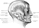
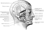
A deep view of the muscles of mastication. The zygoma and masseter muscle are removed.

Head Showing Deep Mastication Muscles

A deep view of the muscles of mastication. The zygoma and masseter muscle are removed.
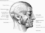
Superficial view of the muscles of mastication.

Head Showing Mastication Muscles

Superficial view of the muscles of mastication.

The pterygoid region.

Head Showing Pterygoid Region

The pterygoid region.

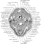
Section of the head through the body of the mandible.

Cross Section of Head Through the Body of the Mandible

Section of the head through the body of the mandible.
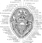
Section of the head through the inferior portion of the mandible.

Cross Section of Head Through the Inferior Portion of the Mandible

Section of the head through the inferior portion of the mandible.
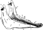
Inferior maxilla of a horse-anterolateral view. Labels: a, body; b, b', rami; c, neck; d, mental foramen; e, buccinator; e', masseter surface; f, f', inner surface of ramus; g, molor alveoli; g', anterior border; g', bar, bounding diastema; h, h', h', posterior border. The angle lies between h' and h'; i, condyles; km coronoid processes; l, maxillary space; m, inferior dental foramen; n, sigmoid notch.

Inferior Maxilla of a Horse

Inferior maxilla of a horse-anterolateral view. Labels: a, body; b, b', rami; c, neck; d, mental foramen;…
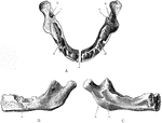
The lower jaw at birth. A, As seen from above. B, As seen from outer side. C, As seen from inner side. Labels: a, mental foramen; b, inferior dental canal; c, lingula; d, sockets fro the dental sacs.

Jaw at Birth

The lower jaw at birth. A, As seen from above. B, As seen from outer side. C, As seen from inner side.…
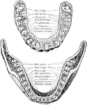
Horizontal section through both the upper and lower jaws to show the roots of the teeth. The sections were carried through the bones a short distance from the edge of their alveolar borders. The upper figure shows the upper teeth, the lower figure the lower teeth. Note the flattened roots of the lower incisors, the two root canals in the anterior root of each lower molar, and the confluence of the three roots of the upper wisdom teeth.

Jaw Showing Roots of Teeth

Horizontal section through both the upper and lower jaws to show the roots of the teeth. The sections…

Inner surface of lower jaw, showing various areas.

Areas of Lower Jaw

Inner surface of lower jaw, showing various areas.

"Appendages of Norway lobster. Ex., Exopodite: En., endopodite; protopodite dark throughout; Ep., epipodite. 1. Antennule--E., position of ear; 2., antenna--K., opening of kidney; 3. mandible--P., palp; 4. first maxilla; 5. second maxilla--B., baler; 6. first maxillipede; 7. second maxillipede; 8. third maxillipede--the basal joint of the protopodite is called coxopodite, the next basipodite; the five joints of the endopodite are called--ischiopodite (i.); meropodite (m.); carpopodite (c.); propodite (p.); dactylopodite (d.); 9. forceps--(7) coxopodite; (6) basipodite, the joints of the endopodite are numbered; 10-13. walking legs; 14. modified male appendage; 15-18. small swimmerets; 19. large paddles." -Thomson, 1916

Norway Lobster Appendages

"Appendages of Norway lobster. Ex., Exopodite: En., endopodite; protopodite dark throughout; Ep., epipodite.…
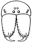
The mandibles of the common garden spider.

Mandible

The mandibles of the common garden spider.

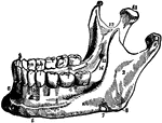
The mandible is the largest and strongest bone of the face. It serves for the reception of the lower teeth. It consists of a curved, horizontal portion, the body, and two perpendicular portions, the rami, which join the back party of the body nearly at right angles.

Mandible

The mandible is the largest and strongest bone of the face. It serves for the reception of the lower…

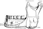
The right half of the mandible.

Mandible

The right half of the mandible.

Lateral half of mandible of a rabbi, opened to show the arrangement of rodent teeth.

Mandible of a Rabbit

Lateral half of mandible of a rabbi, opened to show the arrangement of rodent teeth.

The mandible (lower jaw bone).

The Mandible

The mandible (lower jaw bone).

"The head of female mosquito (culex). a, antenna; c, clypeus; h, hypopharynx; m, mandibles; ma., maxillas; m.p., maxillary palpus; l, labium; la., labrum (epi-pharynx)." — Galloway

Mosquito Head

"The head of female mosquito (culex). a, antenna; c, clypeus; h, hypopharynx; m, mandibles; ma., maxillas;…
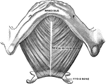
Shown is the mylohyoid muscle. The mylohyoid muscle is a flat, triangular muscle situated beneath the interior belly of the Digastric, forming the muscular floor for the cavity of the mouth.

Mylohyoid Muscle

Shown is the mylohyoid muscle. The mylohyoid muscle is a flat, triangular muscle situated beneath the…

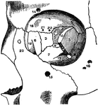
The orbit of the eye.

The Orbit of the Eye

The orbit of the eye.

"Diagrammatic vertical section of the head and proboscis of a mosquito. l, labium bent as when the other parts pierce the skin; p, pumping organ; o, hypopharynx through which is a channel for the exit of the secretion of the poison and salivary glands pn and s; d, exit for poison and saliva; m, maxilla; ma, mandible; la, labrum below which is the channel conducting the food from the inlet at n; st, stomach." —Davison, 1906

Proboscis

"Diagrammatic vertical section of the head and proboscis of a mosquito. l, labium bent as when the other…
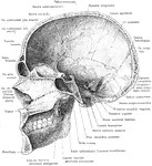
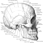
Skull and mandible, lateral view.

Skull and Mandible

Skull and mandible, lateral view.

Median section of the skull and mandible, viewed from the left.

Median Section of the Skull and Mandible

Median section of the skull and mandible, viewed from the left.

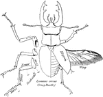
"The largest European species of beetle an adult male sometimes reaching an length of over two inches, and having mandibles about one inch long, which bear some resemblence to the antlers of a stag."—Finley, 1917

Stag Beetle

"The largest European species of beetle an adult male sometimes reaching an length of over two inches,…
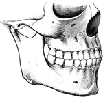
To show the relation of the upper to the lower teeth when the mouth is closed. The manner in which a tooth of one row usually strikes against two teeth of the opposite row, and the resulting interlocking of the teeth, is to be noted.

Teeth

To show the relation of the upper to the lower teeth when the mouth is closed. The manner in which a…

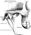
Temporo mandible joint.

Temporo Mandible Joint

Temporo mandible joint.

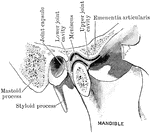
Section through temporo mandible joint.

Section Through Temporo Mandible Joint

Section through temporo mandible joint.

"A typical Thysanuran (Machilis maritima). Female, ventral view. Mx1, Mx2, 1st and 2nd maxillae. ii-x, Appendages on 2nd to 10th abdominal segments. The eversible sacs on the abdominal segments are shown, some protruded and some retracted. Ovp, Ovipositor. Mn, Mandible, and Mxl. maxillula, dissected out of head." — The Encyclopedia Britannica, 1910

Thysanuran

"A typical Thysanuran (Machilis maritima). Female, ventral view. Mx1, Mx2, 1st and 2nd maxillae. ii-x,…