Clipart tagged: ‘"medulla oblongata"’
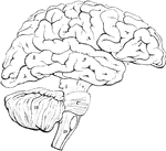
Brain
Diagram illustrating the general relationships of the parts of the brain. Labels: A, fore-brain; b,…
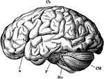
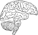
Brain
The brain from the left side. Labels: Cb, the cerebral hemispheres forming the main bulk of the fore-brain;…
Outline of the Encephalon
Plan in outline of the encephalon, as seen from the right side. The parts are represented as separated…
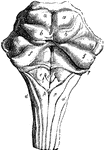
Medulla
Dorsal or posterior view of the medulla, fourth ventricle, and mesencephalon. Labels: p.n., line of…
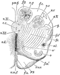
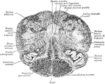
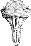
Medulla Oblongata
Anterior or dorsal section of the medulla oblongata in the region of the superior pyramidal decussation.…

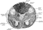
Medulla Oblongata
Section of the medulla oblongata at about the middle of the olivary body. f.l.a., anterior median fissure;…

Anterior Surface of the Pons Varolii and Medulla Oblongata
Ventral or anterior surface of the pons Varolii, and medulla oblongata. Labels: a, anterior pyramids;…
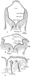
Dorsal Surface of the Pons Varolii
Dorsal or posterior surface of the pons Varolii, corpus quadrigemina, and medulla oblongata. The peduncles…
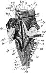
Spinal Cord
Diagrammatic view from before of the spinal cord and medulla oblongata, including the roots of the spinal…