Clipart tagged: ‘mesentery’
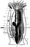
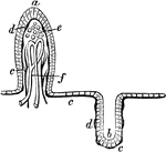
Sea Anemone
"Vertical section of a sea anemone. t., Tentacles; o., mouth; oes., oesophagus; c., c'., apertures through…
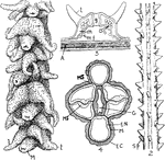
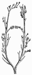
Antipatharians
"Structure of Antipatharians. 1. A group of polyps--M. mouth; t., tentacles. 2. Axis without polyps…
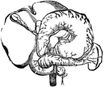
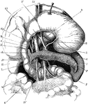
Arteries of the Abdominal Organs
Arteries of the abdominal organs. Labels: 1, the liver; 2, the stomach; 3, upper gut; 4, pancreas; 6,…
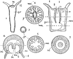
Cnidaria
"Sections of types Coelenterates (diagrammatic): 1 (longitudinal) and 2 (transverse) of a tubular hydroid;…

Coral Shell
"The formation of a coral shell (Asteroides). st., Stomodaeum; ms., mesentery; s., calcareous septum;…
Pacinian Corpuscle
Little, elongated oval bodies, situated on some of the cerebro-spinal and sympathetic nerves.
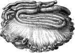
Intestines of an Ox
Mesentery and intestines of an ox. Labels: 1, duodenum; 2, small intestines; 3, caecum; 4, colon; 5,…
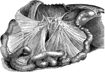
Mesenteries of a Horse
The two mesenteries; the great colon being removed. Labels: a, anterior mesentery; b, mesenteric glands;…
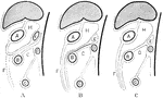
Development of the Mesenteries
Two diagrams to illustrate the development of the mesenteries. In the first figure the rotation of the…
Mucous Membrane
Section of mucous membrane of the small intestine. One the left a villus is seen in seen in section.…
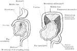
Development of the Great Omentum
Diagram to illustrate the development of the great omentum. A, shows the beginning of the great omentum…
The Stomach, Pancreas, Liver, and Duodenum
The stomach, pancreas, liver, and duodenum, with part of the rest of the small intestine and the mesentery;…
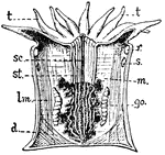
Anthozoan Zooid
"Diagrammtic longitudinal section of an Anthozoan zooid. m, Mesentery; t, Tentacles; st, Stomodaeum;…