There are two fiber types, slow-twitch muscle, or fast-twitch muscle. Most animals have some combination of these two fiber types.

Mustle Fiber

There are two fiber types, slow-twitch muscle, or fast-twitch muscle. Most animals have some combination…

Diagram of muscle fiber with sarcolemma attached. Muscular tissue is the tissue by means of which the active movements of the body are produced. All muscular tissue consists of fibers, and whenever a muscle fiber contracts, it tends to bring its two ends, with whatever may be attached to them, together.

Muscle Fiber

Diagram of muscle fiber with sarcolemma attached. Muscular tissue is the tissue by means of which the…
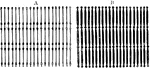
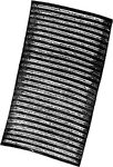
Diagram of the appearance in fresh muscle fiber. Labels: A, At low focus (B) the muscle columns appear dark and in a line with the granules, sarcoplasm light. At high focus (A) the sarcoplasm is dark, muscle columns light, and two rows of granules appear in a line with the sarcoplasm and alternating with the muscle columns.

Muscle Fiber

Diagram of the appearance in fresh muscle fiber. Labels: A, At low focus (B) the muscle columns appear…

Portion of a muscle fiber of Dytiscus, showing network very plainly. One of the transverse networks is split off, and some of the longitudinal bars are shown broken off.

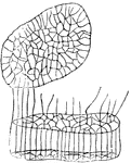
Muscle Fiber Showing Network

Portion of a muscle fiber of Dytiscus, showing network very plainly. One of the transverse networks…

Transverse section of muscle fiber, showing nuclei.

Muscle Fiber Showing Nuclei

Transverse section of muscle fiber, showing nuclei.

Portion of a human muscle fiber.

Portion of a Muscle Fiber

Portion of a human muscle fiber.

Section of a muscle fiber, showing areas of Cohnheim. three nuclei are seen lying close to the sarcolemma.

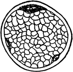
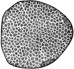
Section of a Muscle Fiber

Section of a muscle fiber, showing areas of Cohnheim. three nuclei are seen lying close to the sarcolemma.

Representation of the direction and arrangement of the fibers in a spindle-shaped muscle (1), a radiated muscle (2). a peniform muscle (3), in a bipeniform muscle (4). Label: t, tendons of a muscle.

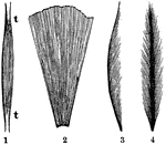
Representation of the Direction and Arrangement of the Muscle Fibers

Representation of the direction and arrangement of the fibers in a spindle-shaped muscle (1), a radiated…

Anastomosing muscle fibers of the heart, seen in longitudinal section. On the right the limits of the separate cells with their nuclei are exhibited somewhat diagrammatically.

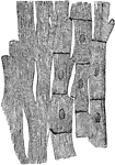
Muscle Fibers of the Heart

Anastomosing muscle fibers of the heart, seen in longitudinal section. On the right the limits of the…

The smooth muscle fibers consist of long spindle-shaped cells, the end of which are frequently spirally twisted, and in the center of which exists a long rod-shaped kernel or nucleus. Unlike striated muscle, they do not form separate muscular masses, but occur scattered, or arranged in more or less dense layers or strata, in almost all organs.

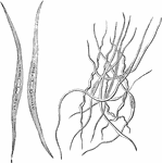
Smooth Muscle Fibers

The smooth muscle fibers consist of long spindle-shaped cells, the end of which are frequently spirally…

The development of muscular fibers from cells. Labels: a, simple cell. b, a pair of cells fused together. c, Three cells fused and their contents assuming the striated character. d, A muscular fiber exhibiting its original composition of cells.

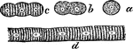
The Development of Muscular Fibers from Cells

The development of muscular fibers from cells. Labels: a, simple cell. b, a pair of cells fused together.…

A fiber of cross-striped muscular tissue, showing the alternating bands.

Fiber of Muscular Tissue Showing Alternating Bands

A fiber of cross-striped muscular tissue, showing the alternating bands.