Clipart tagged: ‘ossification’
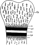
Bone Ossification
Schematic diagram, showing epiphysis and diaphysis and line of ossification. Labels: Ep, epiphysis of…

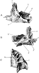
Ossification of the Femur and the Condition of Coxa Vara
Illustrating the ossification of the upper end of the femur and the condition of coxa vara. Labels:…
Ossification of Superior Maxilla
Ossification of superior maxilla. A, outer side. B, inner side. C, under side. Labels: a, nasal process;…
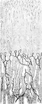
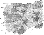
Ossifying Cartilage Showing Calcification
Longitudinal section of ossifying cartilage from the humerus of a fetal sheep. Calcified trabeculae…
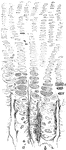
Osteoblasts
Osteoblasts from the parietal bone of a human embryo, thirteen weeks old. Labels: a, bony septa with…