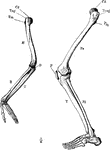
The skeleton of the arm and leg. Labels: H, the humerus; Cd, its articular head which fits into the glenoid fossa of the scapula; U, the ulna; R, the radius; O, the olecranon; Fe, the femur; P, the patella; Fi, the fibula; T, the tibia.

Arm and Leg Skeleton

The skeleton of the arm and leg. Labels: H, the humerus; Cd, its articular head which fits into the…
Bones of the leg

Leg Bones

Bones of the leg

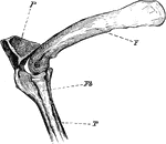
"Phalacrocorax bicristatus. Cormorant. The knee-joint of a Cormorants. F, femur; P, patella; T, tibia; Fb, fibula

The Knee-joint of a Cormorant

"Phalacrocorax bicristatus. Cormorant. The knee-joint of a Cormorants. F, femur; P, patella; T, tibia;…
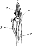
"F. Fibula; T, tibia, with a, its cnemial process, and P, large patella, of a grebe." Elliot Coues, 1884

Leg Bones of a Grebe

"F. Fibula; T, tibia, with a, its cnemial process, and P, large patella, of a grebe." Elliot Coues,…
The knee joint. Labels: h, the patella; f, the tendon.

The Knee Joint

The knee joint. Labels: h, the patella; f, the tendon.

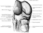
Dissection of knee joint from behind.

Dissection of Knee Joint From Behind

Dissection of knee joint from behind.

Dissection of knee joint from the front with patella thrown down.

Dissection of Knee Joint From Front

Dissection of knee joint from the front with patella thrown down.
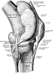
Right knee joint from the lateral surface. The joint cavity and several bursae have been injected with a stiffening medium and then dissected out.

Knee Joint from Lateral Surface

Right knee joint from the lateral surface. The joint cavity and several bursae have been injected with…
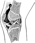
Right knee joint showing interior ligaments.

Knee Joint Showing Interior Ligaments

Right knee joint showing interior ligaments.

Section through right knee joint.

Cross Section Through Knee Joint

Section through right knee joint.

Epiphyseal lines in the neighborhood of the knee joint and their relationship to the synovial membrane.
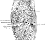
Epiphyseal lines in the Knee Joint

Epiphyseal lines in the neighborhood of the knee joint and their relationship to the synovial membrane.

Frontal section through middle of right knee joint. Seen from behind.

Frontal Section Through Knee Joint

Frontal section through middle of right knee joint. Seen from behind.
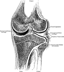
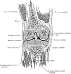
Frontal section through knee joint, showing articulating surfaces and epiphyseal lines.

Frontal Section Through Knee Joint

Frontal section through knee joint, showing articulating surfaces and epiphyseal lines.
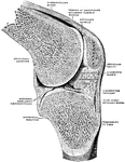
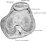
Horizontal section through right knee joint.

Horizontal Section Through Knee Joint

Horizontal section through right knee joint.

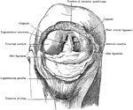
Right knee joint opened, and the knee flexed. Seen from before.

Opened and Flexed Knee Joint

Right knee joint opened, and the knee flexed. Seen from before.

Right knee joint. Sagittal section through the external condyle of the femur. Mesal half of section, from lateral side. The knee is slightly flexed; the joint surfaces have been pulled a little apart.

Sagittal Section Through Knee Joint

Right knee joint. Sagittal section through the external condyle of the femur. Mesal half of section,…
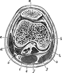
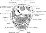
Transverse section of the knee joint through the center of the patella. Labels: a, Bursa patellae; b, internal lateral ligament and inner condyle; c, external lateral ligament and outer condyle; d, biceps; e, semimembranosus; f, semitendinosus; g, gracilis tendon; h, sartorius. 1, internal popliteal nerve; 2, external popliteal nerve; 3, internal saphenous vein.

Transverse Section of the Knee Joint

Transverse section of the knee joint through the center of the patella. Labels: a, Bursa patellae; b,…
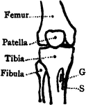
Bones of the knee also showing muscle. Labels: s, insertion of the sartorius; g, insertion of the gracilis.
Bones of the Knee

Bones of the knee also showing muscle. Labels: s, insertion of the sartorius; g, insertion of the gracilis.

Median frontal section of the right knee, viewed from in front.

Median Frontal Section Through Knee

Median frontal section of the right knee, viewed from in front.

Patella removed from right knee, which is strongly flexed to show alar ligaments and ligamentum mucosum. A probe is passed beneath the latter.

Patella Removed from Knee

Patella removed from right knee, which is strongly flexed to show alar ligaments and ligamentum mucosum.…
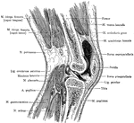
Sagittal section of the right knee, viewed from the outer side. The joint cavity proper lies to each side of the anterior crural ligament.

Sagittal Section Through Knee

Sagittal section of the right knee, viewed from the outer side. The joint cavity proper lies to each…

An anterior view of the right knee-joint.

Knee-joint

An anterior view of the right knee-joint.

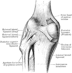
An posterior view of the right knee-joint.

Knee-joint

An posterior view of the right knee-joint.

The right knee-joint showing interior ligaments.

Knee-joint

The right knee-joint showing interior ligaments.

"Bones of the leg. a, femur; b, tibia; c, fibula; d, tarsal bones; e, metatarsal bones; f, phalanges; g, patella." —Martin, 1917

Leg Bones

"Bones of the leg. a, femur; b, tibia; c, fibula; d, tarsal bones; e, metatarsal bones; f, phalanges;…
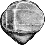
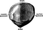
The right patella, dorsal surface.

Patella

The right patella, dorsal surface.

View of the posterior surface of the right patella, showing diagrammatically the areas of contact with the femur in different positions of the knee.

Patella

View of the posterior surface of the right patella, showing diagrammatically the areas of contact with…
Anterior surface (A) and posterior surface (B) of right patella.

Patela

Anterior surface (A) and posterior surface (B) of right patella.

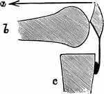
Diagram to show mechanism of fracture of the patella by muscular action. a, Line of action of quadriceps muscle; b, femur; c, tibia.

Mechanism of Fracture of the Patella by Muscular Action

Diagram to show mechanism of fracture of the patella by muscular action. a, Line of action of quadriceps…
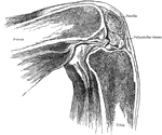
Showing position of patella in relation to condyles of femur with knee flexed at a right angle.

Position of Patella in Relation to Condyles of Femur with Knee Flexed

Showing position of patella in relation to condyles of femur with knee flexed at a right angle.

Showing position of patella in relation to condyles of femur with knee partially flexed.

Position of Patella in Relation to Condyles of Femur with Knee Partially Flexed

Showing position of patella in relation to condyles of femur with knee partially flexed.
Section through the patella and one inch above the condyles.

Cross Section Through Patella

Section through the patella and one inch above the condyles.

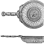
"A round plate or dish. The paterae of the most common kind were small plates of the common red earthenware, on which an ornamental pattern was drawn, and which were sometimes entirely black. The more valuable paterae were metallic, being chiefly of bronze but every family, raised above poverty, possessed one of silver, together with a silver salt-cellar. The preceding cut exhibits a highly ornamented patera, made of bronze. The view of the upper surface is accompanied by a sideview, showing the form and depth of the vessel." — Smith, 1873

Patera

"A round plate or dish. The paterae of the most common kind were small plates of the common red earthenware,…

"The <em>Patella pectonata</em> is found i nthe Mediterranean." &mdash; Goodrich, 1859

Patella pectonata

"The Patella pectonata is found i nthe Mediterranean." — Goodrich, 1859