"Under the power of about 90 diameters the general character of the peridia is seen. They are densely aggregated, elongated, submerged, pale-brown, irregularly torn. The sporidia are copius...Nos. 1 and 2, indicate the general points of growth of this fungus. I find frequently on the leaf-ribs and terminal points of the leaves, and very often dispersed over the smooth parts of the leaf; sometimes, although rarely, the peridia are on the upper surface which, when matured, resembles net work. At the juncture of the leaf (see 4) the cells of the peridia are nearly round, at 5, oblong. From 3 to 4 the cellular structure is of light Vandyke brown; at 5, a pale yellow...6 represents the appearance of the peridia as see by the naked eye; 7, their general arrangements and their groupings on the leaves; 8, three cells, showing the parts of which the peridia are composed being magnified about 125 diameters." -Watts, 1874

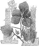
Cellular structure of peridia

"Under the power of about 90 diameters the general character of the peridia is seen. They are densely…

"It is not unusual to fine a decayed spot in the center of potatoes otherwise apparently in good condition. A microscopic examination a portion of the diseased part will show that the decay commenced where the vascular bundles concentrate. At that point the air is in greater volume than elsewhere. Although these mineral acids and caustic alkalies dissolve starch granuals, they do not affect the cellulose cells which contain the starch. When such spots are exposed to the atmosphere the fungus blue-mold forms on the surface. This disease, therefore, has not relation to potato-rot, as ordinarily understood. f represents the mycelium (roots) of blue-mold, Penicillium glaucum, and G that of potato-rot, Peronospora infestans." -Watts, 1874

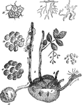
Potato Diseases at a Microscopic Level

"It is not unusual to fine a decayed spot in the center of potatoes otherwise apparently in good condition.…