Diagram of the abdominal part of the alimentary canal (digestive system). Labels: C, the cardiac, and P, the pyloric end of the stomach; D, the duodenum; J, I, the convolutions of the small intestine; CC, the caecum with the vermiform appendix; AC, ascending, TC, transverse, and DC, descending colon; R, the rectum.

Alimentary Canal

Diagram of the abdominal part of the alimentary canal (digestive system). Labels: C, the cardiac, and…

Diagram of the abdominal part of the alimentary canal. Labels: C, the cardiac, and P, the pyloric end of the stomach, A; D, the duodenum; J, I, the convolutions of the small intestine; CC, the caecum with the vermiform appendix; AC, ascending, TC, transverse, and DC, descending colon; SF, sigmoid flexure; R, the rectum.

Alimentary Canal

Diagram of the abdominal part of the alimentary canal. Labels: C, the cardiac, and P, the pyloric end…

The anal cavity and lower part of the rectum in the fetus. Left figure is four month. Middle figure is six months. Right figure is 9 months. In each the anal canal is distinctly marked off from the rectum proper; the columns of Morgagni and the rectal valves are distinct.

Development of the Anal Cavity

The anal cavity and lower part of the rectum in the fetus. Left figure is four month. Middle figure…

The interior of the anal canal and lower part of the rectum, showing the columns of Morgagni and the anal valves between their lower ends. The columns were more numerous in the specimen than usual.

Interior of the Anal Cavity

The interior of the anal canal and lower part of the rectum, showing the columns of Morgagni and the…
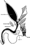
An imperforate anus is the most common congenital defect of the rectum. Shown is a diagram of the termination of the rectum in 54 cases of imperforate anus in the male.

Imperforate Anus

An imperforate anus is the most common congenital defect of the rectum. Shown is a diagram of the termination…
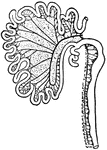
Intestinal tract of <em>Canis vulpes</em>. S, cut end of duodenum; C, caecum; R, cut end of rectum.

Intestinal Tract from Canis Vulpes

Intestinal tract of Canis vulpes. S, cut end of duodenum; C, caecum; R, cut end of rectum.
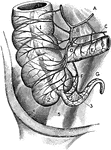
The arterial blood supply of the anterior (ventral) surface of the cecum and appendix. Labels: A, ileocolic artery; B, cecal appendicular artery; D, anterior cecal artery; F and G, Appendicular artery. C, Iliac artery; 1, Right colon; 2, External sacculus of cecum ( to right of taenium coli); 3, Appendix; 4, Iliac muscle; 5, Psoas muscle.

Arterial Blood Supply of the Cecum and Appendix

The arterial blood supply of the anterior (ventral) surface of the cecum and appendix. Labels: A, ileocolic…
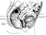
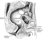
Sagittal section in the median line of the female pelvis.

female Pelvis

Sagittal section in the median line of the female pelvis.

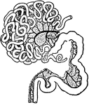
S, cut end of duodenum; R, cut end of rectum; C, vermiform appendix of caecum; X1, X2, X3, cut ends of factors of the portal vein.

Intestinal Tract of a Gorilla

S, cut end of duodenum; R, cut end of rectum; C, vermiform appendix of caecum; X1, X2, X3, cut ends…

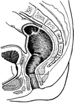
Sagittal section in the median line of the male pelvis.

Male Pelvis

Sagittal section in the median line of the male pelvis.

Diagram of the peritoneum, a serous membrane covering all the contents of the abdominal cavity. Labels: D, diaphragm; L, liver; S, stomach; C, transverse colon; D, transverse duodenum; P, pancreas; I, small intestines; R, rectum; B, bladder.

The Peritoneum

Diagram of the peritoneum, a serous membrane covering all the contents of the abdominal cavity. Labels:…
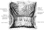
The interior of the anal canal and lower part of the rectum. Showing the column of Morgagni and the anal valves between their lower ends. The columns were more numerous in this specimen than usual.

Rectum and Anal Canal

The interior of the anal canal and lower part of the rectum. Showing the column of Morgagni and the…

The anal canal and rectum in a fetus. A, aged 4 to 5 months; B, 6 moths; C, 9 months. In each he anal canal is distinctly marked off from the rectum proper; the column of Morgagni and the rectal valve are distinct.

Rectum and Anal Canal in Fetus

The anal canal and rectum in a fetus. A, aged 4 to 5 months; B, 6 moths; C, 9 months. In each he anal…

The stages of the rectum and the anal canal on mesial section.

Rectum and Anal Cavity

The stages of the rectum and the anal canal on mesial section.

The blood vessels of the rectum and anus, showing the distribution and anastomosis on the posterior surface near the termination of the gut.

Blood Vessels of the Rectum and Anus

The blood vessels of the rectum and anus, showing the distribution and anastomosis on the posterior…

Inner wall of the lower end of the rectum and anus. On the right the mucous membrane has been removed to show the dilation of the veins and how they pass through the muscular wall to anastomose with the external hemorrhoidal plexus.

Inner Wall of the Rectum and Anus

Inner wall of the lower end of the rectum and anus. On the right the mucous membrane has been removed…
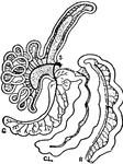
S. cut end of duodenum; R, cut end of rectum; C, caecum; Cl, colon.

Intestinal Tract of a Tapir

S. cut end of duodenum; R, cut end of rectum; C, caecum; Cl, colon.