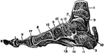
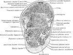
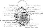
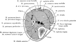
Ankle Joint and Foot
Vertical section of the ankle joint and foot. Labels: 1, tibia; 2, astragalus; 3, os calci; 4, scaphoides;…
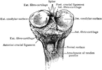
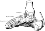
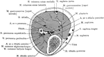
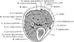
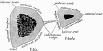
Section Through Ankle Joint
Coronal section through the left ankle joint, astragalus, and calcaneum.
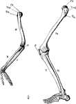
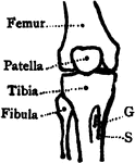
Arm and Leg Skeleton
The skeleton of the arm and leg. Labels: H, the humerus; Cd, its articular head which fits into the…
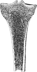
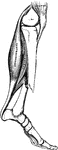
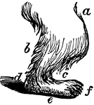
Leg of Bear
This illustration shows the plantigrade leg of a bear. Plantigrade means that the animal walks flat…
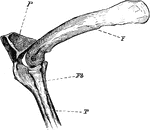
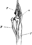
The Knee-joint of a Cormorant
"Phalacrocorax bicristatus. Cormorant. The knee-joint of a Cormorants. F, femur; P, patella; T, tibia;…
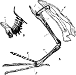
Diver Bones
"A. Pelvis and bones of the leg of the Leon or Diver; i, Innominate bone; f, Thighbone (femur); r, Tibia;…
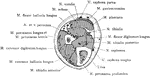
Cross Section Five Inches Above the Lower End of the Fibula
Section five inches above the lower end of the fibula.
Cross Section Four Inches Above the Lower End of the Fibula
Section four inches above the lower end of the fibula.
Leg Bones of a Grebe
"F. Fibula; T, tibia, with a, its cnemial process, and P, large patella, of a grebe." Elliot Coues,…
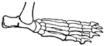
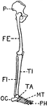
Human Leg (Front View)
This illustration shows a front view of a human leg. P. Pelvis, FE. Femur, TI. Tibia, FI. Fibula, TA.…
Human Leg (Front View), and Comparative Diagrams showing Modifications of the Leg
This illustration shows a human leg (front view), and comparative diagrams showing modifications of…
Human Leg (Side View)
This illustration shows a side view of a human leg. P. Pelvis, FE. Femur, TI. Tibia, FI. Fibula, TA.…
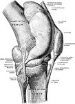
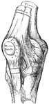
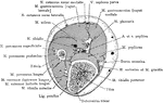
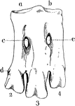
Knee Joint from Lateral Surface
Right knee joint from the lateral surface. The joint cavity and several bursae have been injected with…

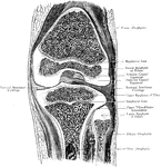
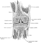
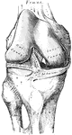
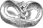
Frontal Section Through Knee Joint
A frontal section through the right knee joint of a boy. Seen from behind.
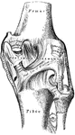
Sagittal Section Through Knee Joint
A sagittal section through the right knee joint of a boy. Seen from the outer side.
Sagittal Section Through Knee Joint
Right knee joint. Sagittal section through the external condyle of the femur. Mesal half of section,…
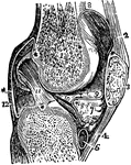
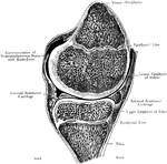
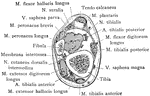
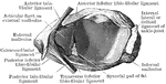
Vertical Section of the Knee Joint
A vertical section of the knee joint. Labels: femur; 3, patella; 2, 4, ligaments of the patella; 5,…
Bones of the Knee
Bones of the knee also showing muscle. Labels: s, insertion of the sartorius; g, insertion of the gracilis.
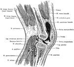
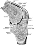
Sagittal Section Through Knee
Sagittal section of the right knee, viewed from the outer side. The joint cavity proper lies to each…
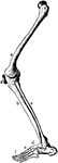
Leg Bones
"Bones of the leg. a, femur; b, tibia; c, fibula; d, tarsal bones; e, metatarsal bones; f, phalanges;…
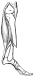
Leg Muscles
The muscles of the front of the leg. 1, tendon of quadriceps; 2, spine tibia; 3, tibialis anticus; 4,…
Cross Section of Leg One Inch Above External Malleolus
Section one inch above the external malleolus.
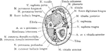
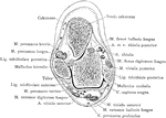
Cross Section Through Leg Three Inches Below Knee Joint
Section through the leg three inches below the right knee joint.
Cross Section of Leg, Two and a Half Inches above Ankle
Section two and a half inches above right ankle joint.
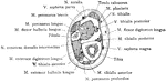
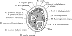
Cross Section Through Leg Two Inches Below Knee Joint
Section through the leg two inches below the right knee joint.
Muscles of the Front of the Leg
Muscles of the front of the leg. Labels: 1, tendon of quadriceps; 2, spine of tibia; 3, tibialis anticus;…
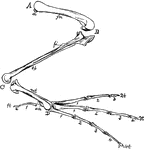
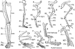
Bones of a Bird's Hind Limb
"Fig 34 - Bones of a bird's hind limb: from a duck, Clangula islandica. A, hip: B, knee: C, heel or…
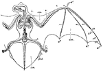
Noctule Bat
"Skeleton and volar Membranes of the Noctule Bat. c, clavicle; h, humerus; r, radius; u, ulna; d1, first…
Mechanism of Fracture of the Patella by Muscular Action
Diagram to show mechanism of fracture of the patella by muscular action. a, Line of action of quadriceps…
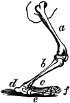
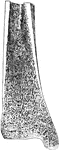
Polar Bear Leg
The polar bear, Plantigrada, is part of the subdivision Carnivora, which includes other carnivorous…
Polar Bear Leg
The anatomy of a polar bear's leg. a, femur (thigh); b, tibia (leg); c, tarsus and metatarsus (foot);…
Shank Bones
A section of the bones of the crus (shank of the leg) taken at about the middle of their length (schematized)…
Front Left Tarsus of a Penguin
An illustration of the front left tarsus of a penguin. "a, articular facet for inner condyle of tibia;…
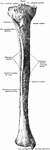
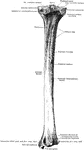
Tibia
Anterior view of the tibia (bone of the leg). Labels: 1, spinous process; 2, surface for condyles of…
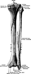
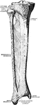
Tibia and Fibula
"The leg consists, like the forearm, of two bones. The larger, a strong, three-sided bone with…
Broken Tibia
"When a bone is broken, blood trickles out between the injured parts, and afterwards gives place to…