Blastoderm of a Chick
| View Cart ⇗ | Info
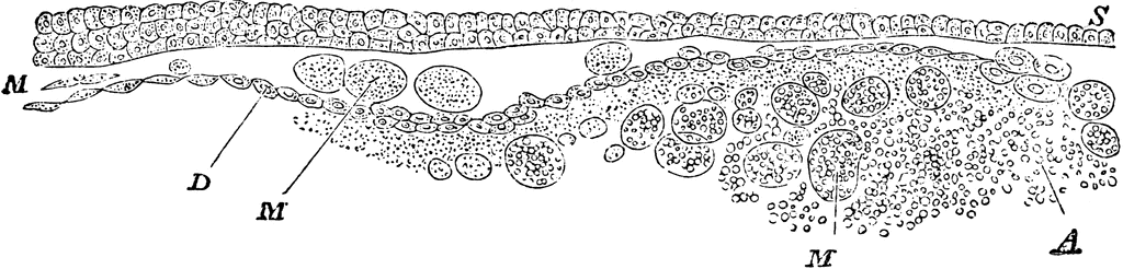
Vertical section of blastoderm of chick (1st day of incubation). S, epiblast consisting of short columnar cells; D, hypoblast, consisting of a single layer of flattened cells; M, formative cells.” They are seen on the right of the figure, passing in between the epiblast and hypoblast to form the mesoblast; A, white yolk granules. Many of the large “formative cells” are seen containing these granules.
Galleries
Bird AnatomySource
Baker, W. Morrant & Harris, Vincent Dormer Kirkes' Hand-book of Physiology, 13th ed. (Philadelphia: P. Blakiston's Son & Co., 1892) 796
Downloads
2400×577, 391.4 KiB
1024×246, 65.4 KiB
640×153, 32.2 KiB
320×76, 9.7 KiB