This human anatomy ClipArt gallery offers 73 illustrations of the human upper respiratory system, including organs involved in respiration. The human upper respiratory tract includes the nasal passages, pharynx, and the larynx.

"To illustrate roughly the passage of air through the glottis, force air through such a tube by blowing hard, and if the strips are not too far apart a sound will be produced. The sound will vary in character as the bands are made tight or loose." — Blaisedell, 1904

Air Passage

"To illustrate roughly the passage of air through the glottis, force air through such a tube by blowing…
The air passages of the lungs.

Air Passages

The air passages of the lungs.

"The air passages of the lungs." — Richardson, 1906

Air Passages

"The air passages of the lungs." — Richardson, 1906

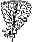
Two alveoli of the lung, highly magnified. Alveoli are cavities which are honeycombed with bulgings in order to increase the surface area exposed to air and covered by capillaries. Labels: b,b, bulgings of the alveoli, a, a.

Two Alveoli of the Lung

Two alveoli of the lung, highly magnified. Alveoli are cavities which are honeycombed with bulgings…
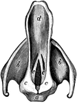
The epiglottis and vocal cords. Labels: e, the vocal cords; d, the epiglottis.

Epiglottis and Vocal Cords

The epiglottis and vocal cords. Labels: e, the vocal cords; d, the epiglottis.
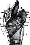
The epiglottis is a cartilaginous lid for the larynx. It is leaf-shaped, situated behind the base of the tongue, and attached by its apex to the posterior surface of the thyroid cartilage, just below the median notch.

The Epiglottis

The epiglottis is a cartilaginous lid for the larynx. It is leaf-shaped, situated behind the base of…
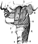
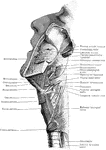
A side view of face. 1 and 2: Trachea. 3: Esophagus. 4, 5, and 6: Muscles. 7: Submaxillary. 8: Parotid gland. 9: Duct from the Parotid gland.

Face

A side view of face. 1 and 2: Trachea. 3: Esophagus. 4, 5, and 6: Muscles. 7: Submaxillary. 8: Parotid…
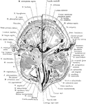
Section of the head immediately below the orbits, at the level of Reid's base line exposing the maxillary sinus.

Cross Section of Head Exposing Maxillary Sinus

Section of the head immediately below the orbits, at the level of Reid's base line exposing the maxillary…
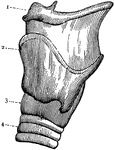
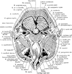
Section of the head through the nasopharynx.

Cross Section of Head Through the Nasopharynx

Section of the head through the nasopharynx.

External view of the left side of Larynx. 1: Front portion of hyoid bone; 2: Upper edge of larynx; 3: Lower portion of larynx; 4: Second ring of trachea.

Larynx

External view of the left side of Larynx. 1: Front portion of hyoid bone; 2: Upper edge of larynx; 3:…
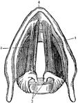
Cross section of the larynx above the vocal cords. 1: Right vocal cord. 2: Left vocal cord. 3: Cartilages to which the vocal cords are attached behind; 4: Front edge of the larynx.

Larynx

Cross section of the larynx above the vocal cords. 1: Right vocal cord. 2: Left vocal cord. 3: Cartilages…
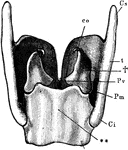
The more important cartilages of the larynx from behind. Labels: t, thyroid; Cs, its superior, and Ci, its inferior, horn of the right side; **, cricoid cartilage; †, arytenoid cartilage; Pv, the corner to which he posterior end of a vocal cord is attached; l'm, corner on which the muscles which approximate or separate the vocal cords are inserted; co, cartilage of Santorini.

Larynx

The more important cartilages of the larynx from behind. Labels: t, thyroid; Cs, its superior, and Ci,…

The larynx viewed from its pharyngeal opening. The back wall of the pharynx has been divided and its edges (11) turned aside. Labels: 1, body of hyoid; 2, its small, and 3, its great, horns; 4, upper and lower horns of thyroid cartilage; 5, mucous membrane of front of pharynx, covering the back of the cricoid cartilage; 6, upper end of gullet; 7, windpipe, lying in front of the gullet; 8, eminence caused by cartilage of Santorini; 9, eminence caused by cartilage of Wrisberg -- both lie in, 10, the aryteno-epiglottidean fold of mucous membrane, surrounding the opening (aditus laryngis) from pharynx to larynx; a, projecting tip of epiglottis; c, the glottis -- the lines leading from the letter point to the free vibrating edges of the vocal cords; b' the ventricles of the larynx -- their upper edges, marking them off from the eminences b, are the false vocal cords.

Larynx

The larynx viewed from its pharyngeal opening. The back wall of the pharynx has been divided and its…
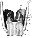
The more important cartilages of the larynx from behind. Labels: t, thyroid; Cs, its superior, and Ci, its inferior, horn of the right side; **, cricoid cartilages;†, arytenoid cartilage; Pv, the corner to which the posterior end of a vocal cord is attached; Pm, corner on which the muscles which approximate or separate the vocal cords are inserted; co, cartilage of Santorini.

Larynx

The more important cartilages of the larynx from behind. Labels: t, thyroid; Cs, its superior, and Ci,…

The larynx viewed from its pharyngeal opening. The back wall of the pharynx has been divided and its edges (11) turned aside. Labels 1, body of hyoid; 2, its small, and 3, its great, horns; 4, upper and lower horns of thyroid cartilages; 5, mucous membrane of front of pharynx, covering the back of the cricoid cartilage; 6, upper end of gullet; 7, windpipe, lying in front of the gullet; 8, eminence caused by cartilage of Santorini; 9, eminence caused by cartilage of Wrisberg; both lie in, 10, the artytenoepiglottic fold of mucous membrane, surrounding the opening (aditus laryngis) from pharynx to larynx; a, projecting tip of epiglottis; c, the glottis, the lines leading from the latter point to the free vibratory edges of the vocal cords; b', the ventricles of the larynx; their upper edges, marking them off from the eminences b, are the false vocal cords.

Larynx

The larynx viewed from its pharyngeal opening. The back wall of the pharynx has been divided and its…
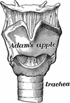
The larynx is made of several pieces of gristle held together by muscle and other tissue. The largest piece of gristle forms the prominence in the throat, called the Adam's apple.

Larynx

The larynx is made of several pieces of gristle held together by muscle and other tissue. The largest…
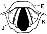
This illustration shows a vertical section of the larynx and its many parts (A. Thyroid Cartilage; B. Cricoid Cartilage; C. hyoid bone; D. Rings of trachea; E. Epiglottis; F. Arytenoid Cartilage; G. Thyro-arytenoid muscle; H. Adam's Apple; I. False Vocal Cords; J. True Vocal Cords; K. Ventricle; L. Rima Glottis).

Vertical Section of Larynx

This illustration shows a vertical section of the larynx and its many parts (A. Thyroid Cartilage; B.…
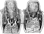
Upper aperture of the larynx in the open (1) and shut (2) position. Labels: A, cushion of epiglottis; B, apices of arytenoids; C, arytenoepiglottidean folds; E, posterior aspect of cricoid; F, false vocal cords; G, rima glottidis,between true vocal cords; H, posterior border of thyroid cartilage; I, tip of great horn of hyoid.

Larynx is Open and Shut Positions

Upper aperture of the larynx in the open (1) and shut (2) position. Labels: A, cushion of epiglottis;…
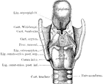
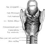
The larynx seen from behind after the removal of the muscles. The cartilages and ligaments only remain.
Larynx Seen from Behind

The larynx seen from behind after the removal of the muscles. The cartilages and ligaments only remain.

The larynx seen from behind to show the intrinsic muscle posteriorly.

Larynx Seen from Behind

The larynx seen from behind to show the intrinsic muscle posteriorly.

Larynx, viewed from above.

Larynx Viewed from Above

Larynx, viewed from above.

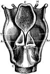
Labels: T, thyroid cartilage: C, cricoid cartilage; Tr, trachea; H, hyoid bone; E, epiglottis; I, joint of thyroid cartilage; Ar, arytenoid cartilages.

Back View of the Larynx

Labels: T, thyroid cartilage: C, cricoid cartilage; Tr, trachea; H, hyoid bone; E, epiglottis; I, joint…

Cavity of larynx, as seen by means of the laryngoscope. A, the rima glottidis closed. B, the rima glottidis widely opened. Labels: 1, rings of the trachea; 2, processus vocalis of the arytenoid cartilage; 3, arytenoepiglottidean fold; 4, sinus pyriformis; 5, laryngeal sinus; 6, vallecula; 7, true vocal cord; 8, cushion of epiglottis; 9, middle glossoepiglottidean fold; 10, base of tongue; 11, epiglottis; 12, false vocal cord; 13, cuneiform tubercle; 14, tubercle of Santorini.

Cavity of Larynx

Cavity of larynx, as seen by means of the laryngoscope. A, the rima glottidis closed. B, the rima glottidis…

Coronal section of the larynx, rear view of front half.

Coronal Section of the Larynx

Coronal section of the larynx, rear view of front half.

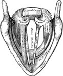
Coronal section through larynx, to show its three compartments.

Coronal Section Through Larynx

Coronal section through larynx, to show its three compartments.

"Cartilages and Ligaments of the Larynx. (Front view.) <em>A</em>, hyoid bone; <em>B</em>, membrane attached to hyoid bone and the shield-shaped cartilage below (thyroid); edges of this shield-shaped cartilage unite at <em>C</em> (Adam's apple is the V-shaped groove on a line with <em>B</em> and <em>C</em>; <em>D</em>, membrane between the shield-shaped cartilage and the signet-ring cartilage below; <em>E</em> cricoid, or signet-ring, cartilage; <em>F</em>, upper ring of the windpipe." &mdash; Blaisedell, 1904

Front view of the larynx

"Cartilages and Ligaments of the Larynx. (Front view.) A, hyoid bone; B, membrane…
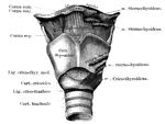
The part of the respiratory tract between the pharyna and the trachea. Responsible for speech.

Human Larynx

The part of the respiratory tract between the pharyna and the trachea. Responsible for speech.
Interior of the larynx, seen from above.

Interior of the Larynx

Interior of the larynx, seen from above.

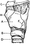
This illustration shows a lateral aspect of the larynx and its multiple parts (A. Thyroid Cartilage; B. Cricoid Cartilage; C. hyoid bone; D. Rings of trachea; E. Epiglottis; F. Arytenoid Cartilage; G. Thyro-arytenoid muscle; H. Adam's Apple; I. False Vocal Cords; J. True Vocal Cords; K. Ventricle; L. Rima Glottis).

Lateral Aspect of Larynx

This illustration shows a lateral aspect of the larynx and its multiple parts (A. Thyroid Cartilage;…

Mesial section through larynx, to show the outer wall of the right half.

Mesial Section Through Larynx

Mesial section through larynx, to show the outer wall of the right half.

"Cartilages and Ligaments of the Larynx. (Front view.) <em>A</em>, epiglottis; <em>B</em>, thyroid cartilage; <em>C</em>, ladle-shaped cartilage; <em>E</em>, cricoid cartilage; <em>F</em>, upper ring of the windpipe." &mdash; Blaisedell, 1904

Posterior view of the larynx

"Cartilages and Ligaments of the Larynx. (Front view.) A, epiglottis; B, thyroid cartilage;…

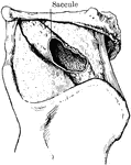
Specimen showing a great extension of the saccule of the larynx.

Saccule of the Larynx

Specimen showing a great extension of the saccule of the larynx.

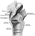
Sagittal section of the larynx, right half.

Sagittal Section of the Larynx

Sagittal section of the larynx, right half.

A section of the larynx. Labels: 1, The trachea. 2, The lower vocal cords. 3, The upper vocal cords. 4, Glottis. 5, The ventricles of the larynx.

Section of the Larynx

A section of the larynx. Labels: 1, The trachea. 2, The lower vocal cords. 3, The upper vocal cords.…

Labels: T, thyroid cartilage: C, cricoid cartilage; Tr, trachea; H, hyoid bone; E, epiglottis; I, joint of thyroid cartilage.

Side View of the Larynx

Labels: T, thyroid cartilage: C, cricoid cartilage; Tr, trachea; H, hyoid bone; E, epiglottis; I, joint…
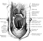
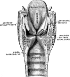
Superior aperture of larynx, exposed by laying open the pharynx from behind.

Superior Aperture of Larynx

Superior aperture of larynx, exposed by laying open the pharynx from behind.
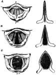
The larynx as seen by means of the laryngoscope in different conditions of the glottis. Labels: A, while singing a high note; b, in quiet breathing; C, during a deep inspiration; l, base of tongue; e, upper free edge of epiglottis; e', cushion of the epiglottis; ph, part of anterior wall of pharynx; cv, the true vocal cords; cvs, the false vocal cords; tr, the trachea with its rings; b, the two bronchi at their commencement.

The Larynx in Different Conditions of the Glottis

The larynx as seen by means of the laryngoscope in different conditions of the glottis. Labels: A, while…

View of the upper part of the larynx as seen by means of the laryngoscope during the utterance of a grave note. Labels: c, epiglottis; s, tubercles of the cartilages of Santorini; a, arytenoid cartilages; z, base of the tongue; ph, the posterior wall of the pharynx.

Upper Part of the Larynx

View of the upper part of the larynx as seen by means of the laryngoscope during the utterance of a…

Front view of cartilages of larynx, trachea and Bronchi.

Front View of the Cartilages of the Larynx, Trachea and Bronchi.

Front view of cartilages of larynx, trachea and Bronchi.
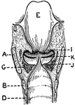
This illustration shows a longitudinal section of  the larynx as seen from behind (A. Thyroid Cartilage; B. Cricoid Cartilage; C. hyoid bone; D. Rings of trachea; E. Epiglottis; F. Arytenoid Cartilage; G. Thyro-arytenoid muscle; H. Adam's Apple; I. False Vocal Cords; J. True Vocal Cords; K. Ventricle; L. Rima Glottis).

Longitudinal Section of Larynx Seen from Behind

This illustration shows a longitudinal section of the larynx as seen from behind (A. Thyroid Cartilage;…
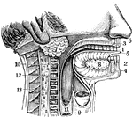
The mouth and neck laid open. 1: The teeth. 3 and 4: Upper and lower jaws. 5: The tongue. 7: Parotid gland. 8: Sublingual gland. 9: Trachea (wind-pipe). 10 and 11: Esophagus (gullet). 12: Spinal column. 13: Spinal cord.

Mouth and Neck

The mouth and neck laid open. 1: The teeth. 3 and 4: Upper and lower jaws. 5: The tongue. 7: Parotid…
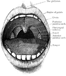
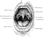
Open mouth showing palate and tonsils. It also shows the two palatine arches, and the pharyngeal isthmus through which the nasopharynx above communicates with the oral portion of the pharynx below.

Mouth Showing Palate and Tonsils

Open mouth showing palate and tonsils. It also shows the two palatine arches, and the pharyngeal isthmus…

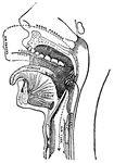
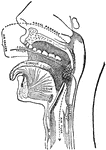
The mouth, nose, and pharynx, with the commencement of gullet (esophagus) and larynx, as exposed by a section a little to the left of the median plane of the head. Labels: a, vertebrae; b, gullet; c, trachea; d, larynx; e, epiglottis; f, soft palate; g, opening of Eustachian tube; k, tongue; l, hard palate; o, p, q, inferior turbinate bones of left nasal chamber.

The Mouth, Nose, and Pharynx

The mouth, nose, and pharynx, with the commencement of gullet (esophagus) and larynx, as exposed by…

The mouth, nose, and pharynx, with the larynx and commencement of gullet (esophagus), seen in section. Labels: a, vertebral column; b, gullet (esophagus); c, trachea; d, larynx; e, epiglottis; f, soft palate, between f and e is the opening at back of cavity or faces; g, opening of Eustachian tube; h, nasal cavity; k, tongue; l, hard palate; m, sphenoid bone at base of skull; n, roof of nasal cavity; o, p, q, placed in nasal cavity.

The Mouth, Nose, and Pharynx

The mouth, nose, and pharynx, with the larynx and commencement of gullet (esophagus), seen in section.…

The mouth, nose, and pharynx, with the commencement of the gullet (esophagus) and larynx, as exposed by a section, a little to the left of the median plane of the head. Labels: a, vertebral column; b, gullet; c, windpipe; d, larynx; e, epiglottis; f, soft palate; g, opening of Esutachian tube; k, tongue; l, hard palate; m; the sphenoid bone on the base of the skull; n, the fore part of the cranial cavity; o,p,q, the turbinate bones of the outer side of the left nostril chamber.

The Mouth, Nose, and Pharynx

The mouth, nose, and pharynx, with the commencement of the gullet (esophagus) and larynx, as exposed…
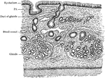
Section of respiratory mucous membrane covering nasal septum.

Respiratory Mucous Membrane

Section of respiratory mucous membrane covering nasal septum.

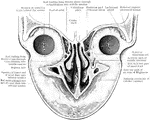
Vertical coronal section through the anterior part of the orbital and nasal cavities and the upper lip.

Section Through Nasal and Orbital Cavities

Vertical coronal section through the anterior part of the orbital and nasal cavities and the upper lip.

"Diagram of a Sectional View of Nasal and Throat Passageways. <em>C</em>, nasal cavities; <em>T</em>, tongue; <em>L</em>, lower jaw; <em>M</em>, Mouth; <em>U</em>, uvula; <em>E</em>, epiglottis; <em>G</em>, larynx; <em>O</em>, gullet, or esophagus." &mdash; Blaisedell, 1904

Nasal and throat passageways

"Diagram of a Sectional View of Nasal and Throat Passageways. C, nasal cavities; T,…
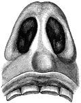
"Nasal Cavities, seen from Below. The sense of smell is located in the membrane which lines the cavities of the nose. This delicate membrane, over which the fibers of the olfactory nerves, or the nerves of smell, are distributed, is kept continually mois by the mucus which it secretes." &mdash; Blaisedell, 1904

Nasal cavities

"Nasal Cavities, seen from Below. The sense of smell is located in the membrane which lines the cavities…
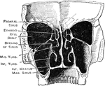
Nasal cavity, right lateral wall, from the left.

Nasal Cavity

Nasal cavity, right lateral wall, from the left.

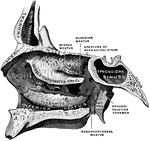
Transverse vertical section of the nasal cavities and accessory sinuses.

Nasal Cavity and Accessory Sinuses

Transverse vertical section of the nasal cavities and accessory sinuses.

Anterior part of section across neck at level of false vocal cords; on left side ventricle of larynx is exposed.

Anterior Part of Section Across Neck at False Vocal Cords

Anterior part of section across neck at level of false vocal cords; on left side ventricle of larynx…

Anterior part of section across neck at level of fourth cervical vertebra, passing through upper part of superior aperture of larynx.

Anterior Part of Section Across Neck at Fourth Cervical Vertebra

Anterior part of section across neck at level of fourth cervical vertebra, passing through upper part…
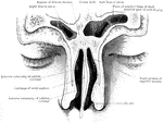
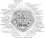
Section of the neck through larynx.

Cross Section of Neck Through the Larynx

Section of the neck through larynx.

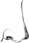
A nose

Nose

A nose

Vertical coronal section through the nose and frontal sinuses.

Section Through Nose and Frontal Sinuses

Vertical coronal section through the nose and frontal sinuses.

The oral cavity seen from in front.

Oral Cavity

The oral cavity seen from in front.

Lateral view of the wall of the pharynx.

Wall of the Pharynx

Lateral view of the wall of the pharynx.

Outline showing the general form of the larynx, trachea, and bronchi, as seen from behind. Labels: h, the great cornu of the hyoid bone; e,epiglottis; t, superior cornu of the thyroid cartilage ; t1, inferior cornu of the thyroid cartilage; a, points to the back of both the arytenoid cartilages, which are surmounted by the cornicula; c, the middle ridge on the back of the cricoid cartilage; tr, the posterior membranous part of the trachea; b, b1, right and left bronchi.

Back View of Respiratory Apparatus

Outline showing the general form of the larynx, trachea, and bronchi, as seen from behind. Labels: h,…

Outline showing the general form of the larynx, trachea, and bronchi, as seen from front. Labels: h, the great cornu of the hyoid bone; e,epiglottis; t, superior and inferior cornu of the thyroid cartilage; c, middle of the cricoid cartilage; tr, the trachea, showing sixteen cartilaginous rings; b, the right bronchus; b1, the left bronchus.

Front View of Respiratory Apparatus

Outline showing the general form of the larynx, trachea, and bronchi, as seen from front. Labels: h,…