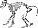
"The skeleton, more especially in the higher forms, is in the main similar to that of man, so that only a brief notice is necessary. In the skull considerable variation in regard to the proportionate length of the face to that of the brain—case (cranial portion) exists in the two sexes, owing to the general development of large tusks in the males." — Encyclopaedia Britannica Company, 1910

Chacma Baboon Skeleton

"The skeleton, more especially in the higher forms, is in the main similar to that of man, so that only…
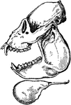
A side view of the howler monkey skull. The monkey have four sharp canines, long teeth on skull, on top and bottom of the jaw.

Side View of Howler Monkey

A side view of the howler monkey skull. The monkey have four sharp canines, long teeth on skull, on…
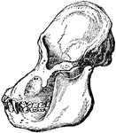
An illustration of an adult male orangutan viewed from the side. The orbit part of the skull is more rounded than humans. The cranium, top part of the skull, and the mouth area are elongated compared to humans.

Adult Male Orangutan Skull Viewed from Side

An illustration of an adult male orangutan viewed from the side. The orbit part of the skull is more…
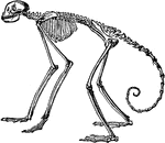
"In the other forms the number (vertebrae) varies between twenty and thirty three, the latter being spider monkeys. The proportion in the spider monkeys of the genus Ateles, almost three to one; in the other longest—tailed genera it is rarely so large as two to one." —Encyclopaedia Britannica, 1910

Side View of Skeleton of South American Spider Monkey

"In the other forms the number (vertebrae) varies between twenty and thirty three, the latter being…

"The greatest absolute length of the fore—limb occurs in the gorilla and the orangutan. The humerus never has a perforation on the inner side of its lower extremity. Except in the man—like apes, the ulna articulates with the wrist (carpus). The hand is capable of pronation and supination on the fore—arm; and except in man, the chimpanzee and the gorilla there is a centrale in the carpus." —Encyclopaedia Britannica, 1910

Front View of Gorilla Skeleton

"The greatest absolute length of the fore—limb occurs in the gorilla and the orangutan. The humerus…
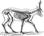
"The bones in the extremities of this the fleetest of quadrupeds are inclined very obliquely towards each other, and towards the scapular and iliac bones. This arrangement increases the leverage of the muscular system and confers great rapidity on the moving parts."—Pettigrew, 1874

Skeleton of the Deer

"The bones in the extremities of this the fleetest of quadrupeds are inclined very obliquely towards…

"Shows how the bones of the arm (a), forearm (b), and hand (c), are twisted, and form a conical screw."—Pettigrew, 1874

Wing of Bird

"Shows how the bones of the arm (a), forearm (b), and hand (c), are twisted, and form a conical screw."—Pettigrew,…
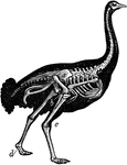
"Shows the powerful legs, small feet, and rudimentary wings of the bird; the obliquity at which the bones of the legs and wings are placed, and the comparatively small angles which two bones make at their point of junction."—Pettigrew, 1857

Skeleton of Ostrich

"Shows the powerful legs, small feet, and rudimentary wings of the bird; the obliquity at which the…
The skeleton of a man.

Human Skeleton

The skeleton of a man.

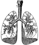
Human lungs. 1 and 2 make up the larynx, or voice box. 1 is thyroid cartilage, 2 is cricoid cartilage. A thin layer is held stretched from the top of the thyroid cartilage and extends to the hyoid bone, which is the only bone in the body that is not connected to another bone.

Human Lungs

Human lungs. 1 and 2 make up the larynx, or voice box. 1 is thyroid cartilage, 2 is cricoid cartilage.…
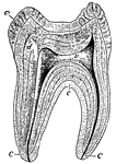
A sectional view of a human molar. The roots, or fangs, are shown covered by a layer of bone called "cement", which is represented by letter c. The center of the tooth is the vascular pulp-cavity v, d is ivory or dentine, and e is the hardest tissue of the body - enamel.

Human Tooth

A sectional view of a human molar. The roots, or fangs, are shown covered by a layer of bone called…
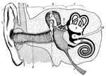
A diagram of the human ear. It is divided into the outer ear - A, middle ear - B, and inner ear - C. 1: ear trumpet or pinna, 2: outer ear passage with the drum that vibrates when sound waves hit it or the tympanum (3) across the inner ear, 4-5-6: the ear ossicles: hammer (malleus) anvil (incus) and stirrup (stapes) which transmit vibrations from the drum to the inner ear (they are also the smallest bones in the body), 7: the Eustachian tube, which connects to the mouth and through which air can indirectly enter, 8: largest chamber in the inner ear, called the utriculus, that has three semicircular canals, 9: the smallest chamber, called the sacculus, which is connected to the cochlea (10) which is the important hearing organ, 11: a "window" in the periotic bone, called the fenestra rotunda, 12: the endolymphatic duct.

Human Ear

A diagram of the human ear. It is divided into the outer ear - A, middle ear - B, and inner ear - C.…

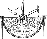
This illustration shows the first stage in the manufacturing of glue. The material used for making the glue (either animal bone or hide) is soaked in a milk of lime (or in some cases, Hydrochloric or Sulfurous acid) to purify and loosen particles.

First Stage in Glue Manufacturing

This illustration shows the first stage in the manufacturing of glue. The material used for making the…
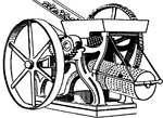
This illustration shows a cross mill and the sieves used to crush and filter bones in glue manufacturing.
Cross Mill and Sieves (Glue)

This illustration shows a cross mill and the sieves used to crush and filter bones in glue manufacturing.

This illustration shows a wash box used for removing lime from materials used in glue manufacturing.

Wash Box for Removing Lime

This illustration shows a wash box used for removing lime from materials used in glue manufacturing.

This illustration shows a section of a human kidney (A, Cortical substance; B, Pyramids; C, Hilum; D, Pelvis; E, Ureter; F, Suprarenal capsule).
Kidneys are excretory organs whose function is to get rid of nitrogenous waste.

Section of Human Kidney

This illustration shows a section of a human kidney (A, Cortical substance; B, Pyramids; C, Hilum; D,…

This illustration shows a section on the knee (A, Femur; B, Tibia; C, Patella; D, Synovial sac; E, bursæ). The knee is a hinge-joint, and the bones entering into its formation are the lower end of the femur, the upper end of the tibia, and the posterior surface of the patella (knee-cap).

Section of the Knee

This illustration shows a section on the knee (A, Femur; B, Tibia; C, Patella; D, Synovial sac; E, bursæ).…
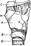
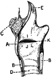
This illustration shows a lateral aspect of the larynx and its multiple parts (A. Thyroid Cartilage; B. Cricoid Cartilage; C. hyoid bone; D. Rings of trachea; E. Epiglottis; F. Arytenoid Cartilage; G. Thyro-arytenoid muscle; H. Adam's Apple; I. False Vocal Cords; J. True Vocal Cords; K. Ventricle; L. Rima Glottis).

Lateral Aspect of Larynx

This illustration shows a lateral aspect of the larynx and its multiple parts (A. Thyroid Cartilage;…
This illustration shows a vertical section of the larynx and its many parts (A. Thyroid Cartilage; B. Cricoid Cartilage; C. hyoid bone; D. Rings of trachea; E. Epiglottis; F. Arytenoid Cartilage; G. Thyro-arytenoid muscle; H. Adam's Apple; I. False Vocal Cords; J. True Vocal Cords; K. Ventricle; L. Rima Glottis).

Vertical Section of Larynx

This illustration shows a vertical section of the larynx and its many parts (A. Thyroid Cartilage; B.…
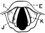
This illustration shows the vocal cords, seen from above during quiet breathing (A. Thyroid Cartilage; B. Cricoid Cartilage; C. hyoid bone; D. Rings of trachea; E. Epiglottis; F. Arytenoid Cartilage; G. Thyro-arytenoid muscle; H. Adam's Apple; I. False Vocal Cords; J. True Vocal Cords; K. Ventricle; L. Rima Glottis).

Vocal Cords Seen from above During Quiet Breathing

This illustration shows the vocal cords, seen from above during quiet breathing (A. Thyroid Cartilage;…
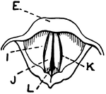
This illustration shows the vocal cords as seen from above during phonation (A. Thyroid Cartilage; B. Cricoid Cartilage; C. hyoid bone; D. Rings of trachea; E. Epiglottis; F. Arytenoid Cartilage; G. Thyro-arytenoid muscle; H. Adam's Apple; I. False Vocal Cords; J. True Vocal Cords; K. Ventricle; L. Rima Glottis).

Vocal Cords Seen from above During Phonation

This illustration shows the vocal cords as seen from above during phonation (A. Thyroid Cartilage; B.…
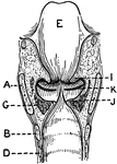
This illustration shows a longitudinal section of  the larynx as seen from behind (A. Thyroid Cartilage; B. Cricoid Cartilage; C. hyoid bone; D. Rings of trachea; E. Epiglottis; F. Arytenoid Cartilage; G. Thyro-arytenoid muscle; H. Adam's Apple; I. False Vocal Cords; J. True Vocal Cords; K. Ventricle; L. Rima Glottis).

Longitudinal Section of Larynx Seen from Behind

This illustration shows a longitudinal section of the larynx as seen from behind (A. Thyroid Cartilage;…

This illustration shows a human leg (front view), and comparative diagrams showing modifications of the leg:
1. Human, 2. Ox, 3. Horse, 4. Bird, 5. Frog, 6. Crocodile, 7. Seal, 8. Dog, 9. Bear
P. Pelvis, FE. Femur, TI. Tibia, FI. Fibula, TA. Tarsus, MT. Metatarsus, PH. Phalanges, OC. Os Calcis.

Human Leg (Front View), and Comparative Diagrams showing Modifications of the Leg

This illustration shows a human leg (front view), and comparative diagrams showing modifications of…
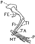
This illustration shows the leg of a crocodile.
P. Pelvis, FE. Femur, TI. Tibia, FI. Fibula, TA. Tarsus, MT. Metatarsus, PH. Phalanges, OC. Os Calcis.

Leg of Crocodile

This illustration shows the leg of a crocodile. P. Pelvis, FE. Femur, TI. Tibia, FI. Fibula, TA. Tarsus,…

This illustration shows the leg of a seal.
P. Pelvis, FE. Femur, TI. Tibia, FI. Fibula, TA. Tarsus, MT. Metatarsus, PH. Phalanges, OC. Os Calcis

Leg of Seal

This illustration shows the leg of a seal. P. Pelvis, FE. Femur, TI. Tibia, FI. Fibula, TA. Tarsus,…
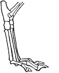
This illustration shows the leg of a dog. This leg is digitigrade. Animals with digitigrade legs walk on their toes (digits).
P. Pelvis, FE. Femur, TI. Tibia, FI. Fibula, TA. Tarsus, MT. Metatarsus, PH. Phalanges, OC. Os Calcis

Leg of Dog

This illustration shows the leg of a dog. This leg is digitigrade. Animals with digitigrade legs walk…

This illustration shows the plantigrade leg of a bear. Plantigrade means that the animal walks flat on its foot.
P. Pelvis, FE. Femur, TI. Tibia, FI. Fibula, TA. Tarsus, MT. Metatarsus, PH. Phalanges, OC. Os Calcis

Leg of Bear

This illustration shows the plantigrade leg of a bear. Plantigrade means that the animal walks flat…
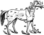
The anatomy of a horse.  1, ears; 2, forelock; 3, forehead; 4, eyes; 5, eye-pits; 6, nose; 7, nostril; 8, point of nose; 9, lips; 10, nether jaw; 11, cheek; 12, poll; 13, mane; 14, withers; 15, parotid glands; 16, throat; 17, neck; 18, jugular vein; 19, shoulder; 20, breast; 21, ribs; 22, back; 23, loins; 24, hip; 25, flank; 26, belly; 27, haunch; 28, thigh; 29, buttock; 30, stifle; 31, leg; 32, tail; 33, hock or hough; 34, cannon or shank bone; 35, arms; 36, knees; 37, passage for girth; 38, elbow; 39, shank; 40, bullet; 41, pasterns; 42, coronet; 43, foot; 44, hoof; 45, fetlock.

Horse

The anatomy of a horse. 1, ears; 2, forelock; 3, forehead; 4, eyes; 5, eye-pits; 6, nose; 7, nostril;…
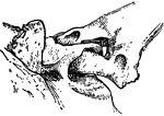
"A joint involving the principle of the shackle. Specifically, in anatomy, a kind of articulation found in the exoskeleton of some fishes, formed by the passing of a bony ring of one part through a perforation of another part, the two being thus movably linked together." —Whitney, 1889
<p>This illustration shows the shackle joint of a large spine with a bony plate of the skin of a siluroid fish.

Shackle Joint from the Exoskeleton of a Siluroid Fish

"A joint involving the principle of the shackle. Specifically, in anatomy, a kind of articulation found…

An illustration of a pichiciago, a small burrowing armadillo. The front half of the animal is covered in the endoskeleton. The back half of the animal is stripped of the outer skeleton so that the exoskeleton or dermoskeleton can be viewed. The scientific name of the pichiciago is <i>Chlamydophorus truncatus</i>.

Armadillo - Endoskeleton and Exoskeleton or Dermoskeleton

An illustration of a pichiciago, a small burrowing armadillo. The front half of the animal is covered…