Internal surface of the bronchi.

Internal Surface of the Bronchi

Internal surface of the bronchi.

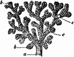
A small bronchial tube. Labels: a, dividing into its terminal branches, c; these have pouched or sacculated walls and end in the sacculated infundibula, b.

A Bronchial Tube

A small bronchial tube. Labels: a, dividing into its terminal branches, c; these have pouched or sacculated…
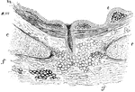
Transverse section of a bronchus. Labels: e, Epithelium (ciliated); immediately beneath it is the mucous membrane or internal fibrous layer, of varying thickness; m, muscular layer; s, m, submucous tissue; f, fibrous tissue; c, cartilage enclosed within the layers of fibrous tissue; g, mucous gland.

Section of the Bronchus

Transverse section of a bronchus. Labels: e, Epithelium (ciliated); immediately beneath it is the mucous…

Front view of cartilages of larynx, trachea and Bronchi.

Front View of the Cartilages of the Larynx, Trachea and Bronchi.

Front view of cartilages of larynx, trachea and Bronchi.

The lungs and air passages seen from the front. On the left of the figure the pulmonary tissue has been dissected away to show the ramifications of the bronchial tubes. Labels: a, larynx; b, trachea; d, right bronchus. The left bronchus is seen entering the root of its lung.

Lungs and Air Passages

The lungs and air passages seen from the front. On the left of the figure the pulmonary tissue has been…

The lungs and air passages seen from the front. On the left of the figure the pulmonary tissue has been dissected away to show the ramifications of the bronchial tubes. Labels: a, larynx; b, trachea; d, right bronchus. The left bronchus is seen entering the root of its lung.

The Lungs and Air Passages

The lungs and air passages seen from the front. On the left of the figure the pulmonary tissue has been…
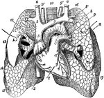
Anterior view of the lungs and heart. Labels: 1, heart; 2, inferior vena cava; 3, superior vena cava; 4, right innominate vein; 5, left innominate vein; 6, jugular vein; 7, subclavian vein; 8, arch of aorta; 8', subclavian artery; 9, left pulmonary artery; 9', 9', carotid artery; 10, trachea; 11, left bronchus; 12, ramifications of right bronchus exposed in upper lobe of right lung; 13, 14, middle lobe; 15, lower lobe; 16, upper lobe of left lung; 17, lower lobe of left lung.

Anterior View of the Lungs and Heart

Anterior view of the lungs and heart. Labels: 1, heart; 2, inferior vena cava; 3, superior vena cava;…

Mediastinal surfaces of the two lungs of a subject hardened by formalin injection. A, right lung. B, left lung. Labels: a, base; 2, fissure; 3, cardiac depression; 4, groove for innominate vein; 5, groove for innominate artery; 6, apex pulmonis; 7, groove for vena azygos major; 8, eparterial bronchus; 9, pulmonary artery (right); 10, fissure; 11, groove for aorta; 12, bronchus; 13, pulmonary artery (left); 14, apex pulmonis; 15, groove for left subclavian artery; 16, groove for left innominate vein; 17, cardiac depression; 18, fissure; 19, base.

Mediastinal Surfaces of the Lungs

Mediastinal surfaces of the two lungs of a subject hardened by formalin injection. A, right lung. B,…
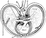
"Cross-section of thorax. A, bronchus, entering the lung; B, the aorta cut at its origin and again at the descending part of its arch; C, the pericardial space; D, the pleural cavity; E, the alimentary canal; PA, the pulmonary artery; X, the neural canal." —Martin, 1917

Thorax

"Cross-section of thorax. A, bronchus, entering the lung; B, the aorta cut at its origin and again at…

The trachea and bronchi. The thyroid body is indicated by a dotted line.

Trachea and Bronchi

The trachea and bronchi. The thyroid body is indicated by a dotted line.