Clipart tagged: ‘caecum’
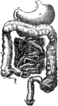
Alimentary Canal
Diagram of the abdominal part of the alimentary canal (digestive system). Labels: C, the cardiac, and…
Vermiform appendix
"A, a portion of the colon laid open to show the valve between the large and small intestine;…
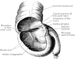
Caecum
Caecum showing ileocaecal valve. The caecum bas been distended with air and dried, and a portion of…
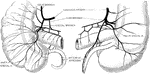
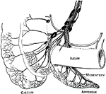
Blood Supply of Caecum and Appendix
The blood supply of the caecum and vermiform appendix. The illustration to the left gives a front view,…
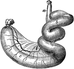
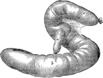
Caecum and Colon of a Dog
Caecum and colon of a dog-inflated. Labels: a, ileum; b, caecum; c, colon.
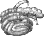
Caecum and Colon of a Hog
Caecum and colon of a hog-inflated. Labels: a, ileum; b, caecum; c, colon; d, rectum.
Caecum and Colon of Horse
Caecum and great colon of a horse. Labels: a, caecum; b, c, its muscular bands; d, termination of the…
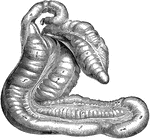
Caecum of an Ox
Caecum and origin of colon of an ox- inflated. Labels: a, terminal portion of the ileum; b, caecum;…
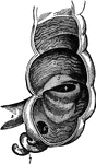
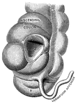
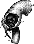
Caecum of the Large Intestine
Caecum, showing its appendix, entrance of ileum, and ileo-caecal valve. The caecum is a large blind…
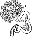
Intestinal Tract from Canis Vulpes
Intestinal tract of Canis vulpes. S, cut end of duodenum; C, caecum; R, cut end of rectum.
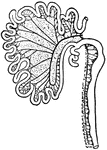
Intestinal Tract of a Gorilla
S, cut end of duodenum; R, cut end of rectum; C, vermiform appendix of caecum; X1, X2, X3, cut ends…
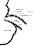
Ileo-caecal valve
The ileo-caecal valve, where the small intestine joins the large. Labels: a, ileum; b, ascending colon;…
Formation of Ileo-caecal Valve
Diagrammatic Section through the junction of the ileum with caecum, to show the formation of the ileocaecal…
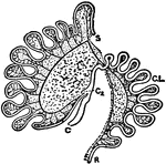
Intestinal Tract of Macropus Bennetti
S, cut end of duodenum; R, cut end of rectum; C, caecum; C2, accessory caecum; C.L., colic loop of hind-gut.